Explore
 Validate
 Learn
Learn Western blot
 Immunoprecipitation
Antibody data
- Antibody Data
- Antigen structure
- References [8]
- Comments [0]
- Validations
- Western blot [2]
- Immunocytochemistry [1]
Submit
Validation data
Reference
Comment
Report error
- Product number
- ABIN152485 - Provider product page

- Provider
- antibodies-online
- Product name
- anti-Internexin Neuronal Intermediate Filament Protein, alpha (INA) antibody
- Antibody type
- Polyclonal
- Description
- Unpurified
- Reactivity
- Human, Mouse, Rat
- Host
- Rabbit
- Vial size
- 0.1 mL
- Storage
- Store at 4°C short term. Aliquot and store at -20°C long term. Avoid freeze-thaw cycles.
- Handling
- Avoid freeze-thaw cycles
Submitted references Congenital Anophthalmia and Binocular Neonatal Enucleation Differently Affect the Proteome of Primary and Secondary Visual Cortices in Mice.
Cortical murine neurons lacking the neurofilament light chain protein have an attenuated response to injury in vitro.
Cytoskeletal changes during development and aging in the cortex of neurofilament light protein knockout mice.
Degeneration of axons in spinal white matter in G93A mSOD1 mouse characterized by NFL and α-internexin immunoreactivity.
Neuron-glia interactions underlie ALS-like axonal cytoskeletal pathology.
Focal damage to the adult rat neocortex induces wound healing accompanied by axonal sprouting and dendritic structural plasticity.
Excitotoxicity mediated by non-NMDA receptors causes distal axonopathy in long-term cultured spinal motor neurons.
Investigation of general and cytoskeletal markers to estimate numbers and proportions of neurons in the human intestine.
Laramée ME, Smolders K, Hu TT, Bronchti G, Boire D, Arckens L
PloS one 2016;11(7):e0159320
PloS one 2016;11(7):e0159320
Cortical murine neurons lacking the neurofilament light chain protein have an attenuated response to injury in vitro.
Blizzard CA, King AE, Vickers J, Dickson T
Journal of neurotrauma 2013 Nov 15;30(22):1908-18
Journal of neurotrauma 2013 Nov 15;30(22):1908-18
Cytoskeletal changes during development and aging in the cortex of neurofilament light protein knockout mice.
Liu Y, Staal JA, Canty AJ, Kirkcaldie MT, King AE, Bibari O, Mitew ST, Dickson TC, Vickers JC
The Journal of comparative neurology 2013 Jun 1;521(8):1817-27
The Journal of comparative neurology 2013 Jun 1;521(8):1817-27
Degeneration of axons in spinal white matter in G93A mSOD1 mouse characterized by NFL and α-internexin immunoreactivity.
King AE, Blizzard CA, Southam KA, Vickers JC, Dickson TC
Brain research 2012 Jul 17;1465:90-100
Brain research 2012 Jul 17;1465:90-100
Neuron-glia interactions underlie ALS-like axonal cytoskeletal pathology.
King AE, Dickson TC, Blizzard CA, Woodhouse A, Foster SS, Chung RS, Vickers JC
Neurobiology of aging 2011 Mar;32(3):459-69
Neurobiology of aging 2011 Mar;32(3):459-69
Focal damage to the adult rat neocortex induces wound healing accompanied by axonal sprouting and dendritic structural plasticity.
Blizzard CA, Chuckowree JA, King AE, Hosie KA, McCormack GH, Chapman JA, Vickers JC, Dickson TC
Cerebral cortex (New York, N.Y. : 1991) 2011 Feb;21(2):281-91
Cerebral cortex (New York, N.Y. : 1991) 2011 Feb;21(2):281-91
Excitotoxicity mediated by non-NMDA receptors causes distal axonopathy in long-term cultured spinal motor neurons.
King AE, Dickson TC, Blizzard CA, Foster SS, Chung RS, West AK, Chuah MI, Vickers JC
The European journal of neuroscience 2007 Oct;26(8):2151-9
The European journal of neuroscience 2007 Oct;26(8):2151-9
Investigation of general and cytoskeletal markers to estimate numbers and proportions of neurons in the human intestine.
Ganns D, Schrödl F, Neuhuber W, Brehmer A
Histology and histopathology 2006 Jan;21(1):41-51
Histology and histopathology 2006 Jan;21(1):41-51
No comments: Submit comment
Supportive validation
- Submitted by
- antibodies-online (provider)
- Main image
- Experimental details
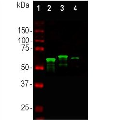
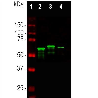
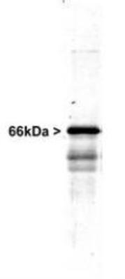
- WB
- Submitted by
- antibodies-online (provider)
- Main image

- Experimental details
- WB
Supportive validation
- Submitted by
- antibodies-online (provider)
- Main image

- Experimental details
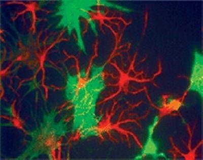
- Polyclonal alpha internexin, ABIN152485 (red) staining neuronal progenitor cells. The green stain shows the fibroblast marker, Plectin (not one of our antibodies).