Explore
 Validate
 Learn
Learn Western blot
 Immunocytochemistry
Antibody data
- Antibody Data
- Antigen structure
- References [2]
- Comments [0]
- Validations
- Western blot [6]
- Immunohistochemistry [18]
Submit
Validation data
Reference
Comment
Report error
- Product number
- NBP1-88221 - Provider product page

- Provider
- Novus Biologicals
- Proper citation
- Novus Cat#NBP1-88221, RRID:AB_11052501
- Product name
- Rabbit Polyclonal Calretinin Antibody
- Antibody type
- Polyclonal
- Description
- Immunogen affinity purified. Specificity of human Calretinin antibody verified on a Protein Array containing target protein plus 383 other non-specific proteins.
- Reactivity
- Human, Mouse
- Host
- Rabbit
- Isotype
- IgG
- Vial size
- 0.1 ml
- Storage
- Store at 4C short term. Aliquot and store at -20C long term. Avoid freeze-thaw cycles.
Submitted references Off-Target Effects in Transgenic Mice: Characterization of Dopamine Transporter (DAT)-Cre Transgenic Mouse Lines Exposes Multiple Non-Dopaminergic Neuronal Clusters Available for Selective Targeting within Limbic Neurocircuitry.
Tissue profiling of the mammalian central nervous system using human antibody-based proteomics.
Papathanou M, Dumas S, Pettersson H, Olson L, Wallén-Mackenzie Å
eNeuro 2019 Sep Oct;6(5)
eNeuro 2019 Sep Oct;6(5)
Tissue profiling of the mammalian central nervous system using human antibody-based proteomics.
Mulder J, Björling E, Jonasson K, Wernérus H, Hober S, Hökfelt T, Uhlén M
Molecular & cellular proteomics : MCP 2009 Jul;8(7):1612-22
Molecular & cellular proteomics : MCP 2009 Jul;8(7):1612-22
No comments: Submit comment
Supportive validation
- Submitted by
- Novus Biologicals (provider)
- Main image
- Experimental details
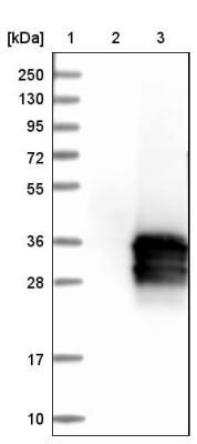
- Western Blot: Calretinin Antibody [NBP1-88221] - Lane 1: Marker [kDa] 250, 130, 95, 72, 55, 36, 28, 17, 10. Lane 2: Negative control (vector only transfected HEK293T lysate). Lane 3: Over-expression lysate (Co-expressed with a C-terminal myc-DDK tag (3.1 kDa) in mammalian HEK293T cells).
- Submitted by
- Novus Biologicals (provider)
- Main image
- Experimental details
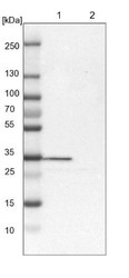
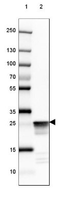
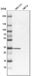
- Western Blot: Calretinin Antibody [NBP1-88221] - Lane 1: NIH-3T3 cell lysate (Mouse embryonic fibroblast cells) Lane 2: NBT-II cell lysate (Rat Wistar bladder tumour cells)

- Submitted by
- Novus Biologicals (provider)
- Main image
- Experimental details
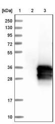
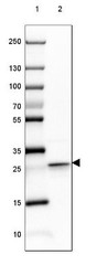
- Western Blot: Calretinin Antibody [NBP1-88221] - Lane 1: Marker [kDa] 250, 130, 100, 70, 55, 35, 25, 15, 10 Lane 2: Human Cerebral Cortex tissue

- Submitted by
- Novus Biologicals (provider)
- Main image
- Experimental details
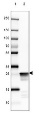
- Western Blot: Calretinin Antibody [NBP1-88221] - Lane 1: Marker [kDa] 250, 130, 100, 70, 55, 35, 25, 15, 10 Lane 2: Mouse Cerebellum tissue
- Submitted by
- Novus Biologicals (provider)
- Main image

- Experimental details
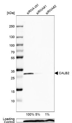
- Western Blot: Calretinin Antibody [NBP1-88221] - Analysis in U2OS cells transfected with control siRNA, target specific siRNA probe #1 and #2,. Remaining relative intensity is presented. Loading control: Anti-PPIB.
- Submitted by
- Novus Biologicals (provider)
- Main image
- Experimental details
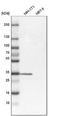
- Western Blot: Calretinin Antibody [NBP1-88221] - Analysis in mouse cell line NIH-3T3 and rat cell line NBT-II.
Supportive validation
- Submitted by
- Novus Biologicals (provider)
- Main image
- Experimental details
- Immunohistochemistry-Paraffin: Calretinin Antibody [NBP1-88221] - Staining of human cerebral cortex shows strong cytoplasmic immunoreactivity in a subset of neurons.
- Submitted by
- Novus Biologicals (provider)
- Main image
- Experimental details
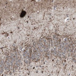
- Immunohistochemistry-Paraffin: Calretinin Antibody [NBP1-88221] - Staining of human hippocampus shows strong cytoplasmic positivity in s subset of neurons.

- Submitted by
- Novus Biologicals (provider)
- Main image

- Experimental details
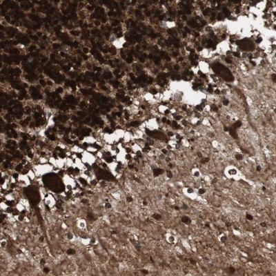
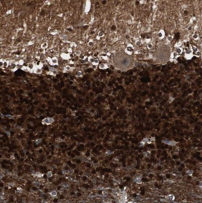
- Immunohistochemistry-Paraffin: Calretinin Antibody [NBP1-88221] - Staining of human cerebellum shows strong cytoplasmic and nuclear positivity in Purkinje cells and strong nuclear positivity in cells in molecular layer.
- Submitted by
- Novus Biologicals (provider)
- Main image
- Experimental details
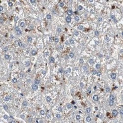
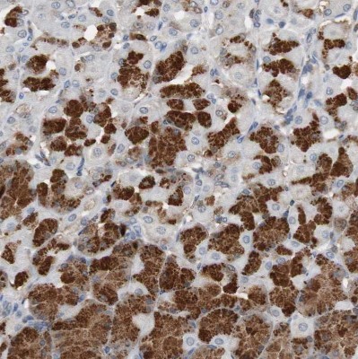
- Immunohistochemistry-Paraffin: Calretinin Antibody [NBP1-88221] - Staining of human liver.

- Submitted by
- Novus Biologicals (provider)
- Main image

- Experimental details
- Immunohistochemistry-Paraffin: Calretinin Antibody [NBP1-88221] - Staining of human soft tissues.

- Submitted by
- Novus Biologicals (provider)
- Main image
- Experimental details
- Immunohistochemistry-Paraffin: Calretinin Antibody [NBP1-88221] - Staining of human cerebral cortex.

- Submitted by
- Novus Biologicals (provider)
- Main image

- Experimental details
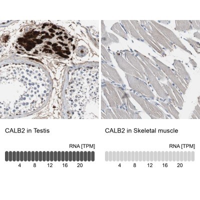
- Immunohistochemistry-Paraffin: Calretinin Antibody [NBP1-88221] - Staining of human testis.

- Submitted by
- Novus Biologicals (provider)
- Main image
- Experimental details
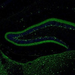
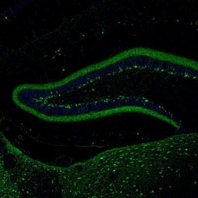
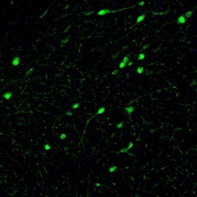
- Immunohistochemistry: Calretinin Antibody [NBP1-88221] - Staining of mouse hippocampus shows selective immunoreactivity in the dentate gyrus neurons.
- Submitted by
- Novus Biologicals (provider)
- Main image
- Experimental details
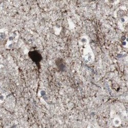
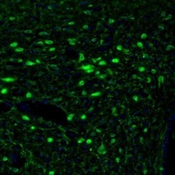
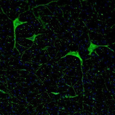
- Immunohistochemistry: Calretinin Antibody [NBP1-88221] - Staining of mouse hypothalamus shows immunoreactivity in neurons of the paraventricular nucleus.

- Submitted by
- Novus Biologicals (provider)
- Main image
- Experimental details
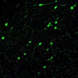
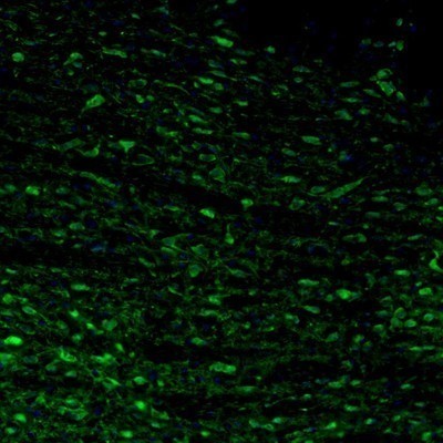
- Immunohistochemistry: Calretinin Antibody [NBP1-88221] - Staining of mouse substantia nigra shows neuronal cell bodies and processes in pars compacta.
- Submitted by
- Novus Biologicals (provider)
- Main image

- Experimental details
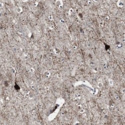
- Immunohistochemistry: Calretinin Antibody [NBP1-88221] - Staining of mouse hindbrain shows cytoplasmic immunoreactivity in neurons of the pontine nucleus.
- Submitted by
- Novus Biologicals (provider)
- Main image

- Experimental details
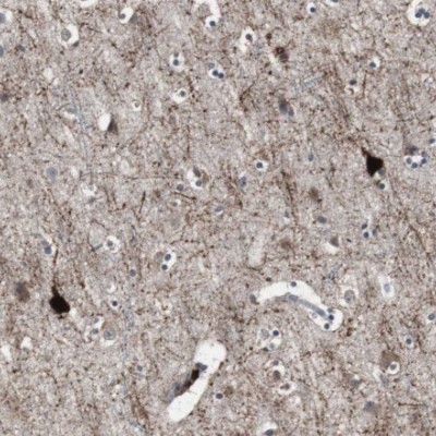
- Immunohistochemistry: Calretinin Antibody [NBP1-88221] - Staining of mouse medulla shows positivity in the gigantocellular reticular nucleus neurons.
- Submitted by
- Novus Biologicals (provider)
- Main image

- Experimental details
- Immunohistochemistry-Paraffin: Calretinin Antibody [NBP1-88221] - Staining of human cerebellum shows strong cytoplasmic and nuclear positivity in cells in granular layer.
- Submitted by
- Novus Biologicals (provider)
- Main image
- Experimental details
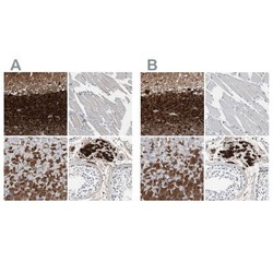
- Immunohistochemistry-Paraffin: Calretinin Antibody [NBP1-88221] - Staining of human cerebellum, skeletal muscle, stomach and testis using Anti-CALB2 antibody NBP1-88221 (A) shows similar protein distribution across tissues to independent antibody NBP1-88220 (B).

- Submitted by
- Novus Biologicals (provider)
- Main image
- Experimental details
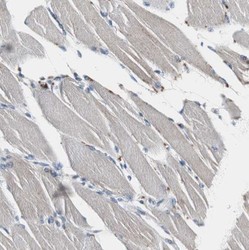
- Immunohistochemistry-Paraffin: Calretinin Antibody [NBP1-88221] - Staining of human skeletal muscle shows no positivity in myocytes as expected.
- Submitted by
- Novus Biologicals (provider)
- Main image
- Experimental details
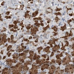
- Immunohistochemistry-Paraffin: Calretinin Antibody [NBP1-88221] - Staining of human stomach shows strong cytoplasmic positivity in glandular cells.
- Submitted by
- Novus Biologicals (provider)
- Main image

- Experimental details
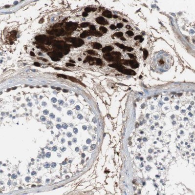
- Immunohistochemistry-Paraffin: Calretinin Antibody [NBP1-88221] - Staining of human testis shows strong cytoplasmic and nuclear positivity in Leydig cells.
- Submitted by
- Novus Biologicals (provider)
- Main image

- Experimental details
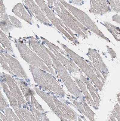
- Immunohistochemistry-Paraffin: Calretinin Antibody [NBP1-88221] - Staining in human testis and skeletal muscle tissues using NBP1-88221 antibody. Corresponding CALB2 RNA-seq data are presented for the same tissues.