 Explore
 Validate
 Learn
Learn Western blot
 Immunohistochemistry
Antibody data
- Antibody Data
- Antigen structure
- References [1]
- Comments [0]
- Validations
- Western blot [6]
- Immunocytochemistry [2]
- Immunohistochemistry [24]
Submit
Validation data
Reference
Comment
Report error
- Product number
- HPA007305 - Provider product page

- Provider
- Atlas Antibodies
- Proper citation
- Atlas Antibodies Cat#HPA007305, RRID:AB_1078386
- Product name
- Anti-CALB2
- Antibody type
- Polyclonal
- Description
- Polyclonal Antibody against Human CALB2, Gene description: calbindin 2, Alternative Gene Names: CAL2, Validated applications: ICC, IHC, WB, Uniprot ID: P22676, Storage: Store at +4°C for short term storage. Long time storage is recommended at -20°C.
- Reactivity
- Human
- Host
- Rabbit
- Conjugate
- Unconjugated
- Isotype
- IgG
- Vial size
- 100 µl
- Concentration
- 0.55 mg/ml
- Storage
- Store at +4°C for short term storage. Long time storage is recommended at -20°C.
Submitted references Tissue Profiling of the Mammalian Central Nervous System Using Human Antibody-based Proteomics
Mulder J, Bjorling E, Jonasson K, Wernerus H, Hober S, Hokfelt T, Uhlen M
Molecular & Cellular Proteomics 2009 July;8(7):1612-1622
Molecular & Cellular Proteomics 2009 July;8(7):1612-1622
No comments: Submit comment
Supportive validation
- Submitted by
- Atlas Antibodies (provider)
- Enhanced method
- Genetic validation
- Main image

- Experimental details
- Western blot analysis in U2OS cells transfected with control siRNA, target specific siRNA probe #1 and #2, using Anti-CALB2 antibody. Remaining relative intensity is presented. Loading control: Anti-PPIB.

- Submitted by
- Atlas Antibodies (provider)
- Main image

- Experimental details
- Lane 1: NIH-3T3 cell lysate (Mouse embryonic fibroblast cells)Lane 2: NBT-II cell lysate (Rat Wistar bladder tumour cells)

- Submitted by
- Atlas Antibodies (provider)
- Main image

- Experimental details
- Lane 1: Marker [kDa] 250, 130, 95, 72, 55, 36, 28, 17, 10Lane 2: Negative control (vector only transfected HEK293T lysate)Lane 3: Over-expression lysate (Co-expressed with a C-terminal myc-DDK tag (~3.1 kDa) in mammalian HEK293T cells, LY400659)

- Submitted by
- Atlas Antibodies (provider)
- Main image

- Experimental details
- Lane 1: Marker [kDa] 250, 130, 100, 70, 55, 35, 25, 15, 10Lane 2: Human Cerebral Cortex tissue

- Submitted by
- Atlas Antibodies (provider)
- Main image

- Experimental details
- Lane 1: Marker [kDa] 250, 130, 100, 70, 55, 35, 25, 15, 10Lane 2: Mouse Cerebellum tissue

- Submitted by
- Atlas Antibodies (provider)
- Main image

- Experimental details
- Western blot analysis in mouse cell line NIH-3T3 and rat cell line NBT-II.

Supportive validation
- Submitted by
- Atlas Antibodies (provider)
- Main image

- Experimental details
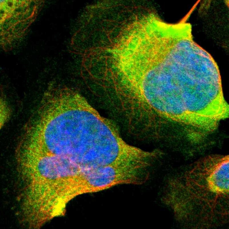
- Immunofluorescent staining of human cell line U-2 OS shows localization to cytosol.
- Sample type
- HUMAN

- Submitted by
- Atlas Antibodies (provider)
- Main image

- Experimental details
- Immunofluorescent staining of human cell line U-2 OS shows localization to cytosol.
- Sample type
- Human
- Protocol
- Protocol
Enhanced validation
Enhanced validation
Enhanced validation
Supportive validation
- Submitted by
- Atlas Antibodies (provider)
- Enhanced method
- Orthogonal validation
- Main image

- Experimental details
- Immunohistochemistry analysis in human testis and skeletal muscle tissues using HPA007305 antibody. Corresponding CALB2 RNA-seq data are presented for the same tissues.
- Sample type
- Human
- Protocol
- Protocol

Enhanced validation
- Submitted by
- Atlas Antibodies (provider)
- Enhanced method
- Independent antibody validation
- Main image

- Experimental details
- Immunohistochemical staining of human cerebral cortex, liver, soft tissues and testis using Anti-CALB2 antibody HPA007305 (A) shows similar protein distribution across tissues to independent antibody HPA007306 (B).

Enhanced validation
- Submitted by
- Atlas Antibodies (provider)
- Enhanced method
- Independent antibody validation
- Main image

- Experimental details
- Immunohistochemical staining of human cerebellum, skeletal muscle, stomach and testis using Anti-CALB2 antibody HPA007305 (A) shows similar protein distribution across tissues to independent antibody HPA007306 (B).
- Sample type
- Human
- Secondary Ab
- Secondary Ab
- Protocol
- Protocol

Supportive validation
- Submitted by
- Atlas Antibodies (provider)
- Main image

- Experimental details
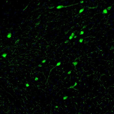
- Immunohistochemical staining of human cerebellum shows strong cytoplasmic and nuclear positivity in Purkinje cells and strong nuclear positivity in cells in molecular layer.

- Submitted by
- Atlas Antibodies (provider)
- Main image

- Experimental details
- Immunofluorescence staining of mouse hippocampus shows selective immunoreactivity in the dentate gyrus neurons.
- Sample type
- HUMAN

- Submitted by
- Atlas Antibodies (provider)
- Main image

- Experimental details
- Immunohistochemical staining of human hippocampus shows strong cytoplasmic positivity in s subset of neurons.

- Submitted by
- Atlas Antibodies (provider)
- Main image

- Experimental details
- Immunofluorescence staining of mouse hypothalamus shows immunoreactivity in neurons of the paraventricular nucleus.
- Sample type
- HUMAN

- Submitted by
- Atlas Antibodies (provider)
- Main image

- Experimental details
- Immunofluorescence staining of mouse substantia nigra shows neuronal cell bodies and processes in pars compacta.
- Sample type
- HUMAN

- Submitted by
- Atlas Antibodies (provider)
- Main image

- Experimental details
- Immunofluorescence staining of mouse hindbrain shows cytoplasmic immunoreactivity in neurons of the pontine nucleus.
- Sample type
- HUMAN

- Submitted by
- Atlas Antibodies (provider)
- Main image

- Experimental details
- Immunofluorescence staining of mouse medulla shows positivity in the gigantocellular reticular nucleus neurons.
- Sample type
- HUMAN

- Submitted by
- Atlas Antibodies (provider)
- Main image

- Experimental details
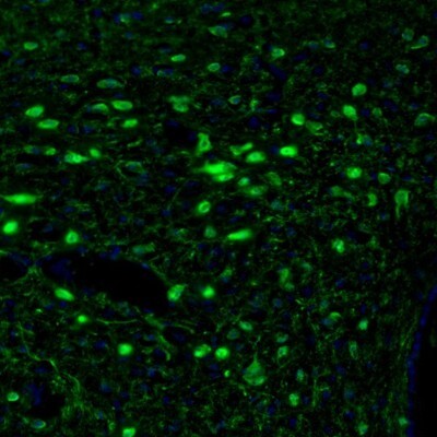
- Immunohistochemical staining of human cerebral cortex shows strong cytoplasmic immunoreactivity in a subset of neurons.

- Submitted by
- Atlas Antibodies (provider)
- Main image

- Experimental details
- Immunohistochemical staining of human testis using Anti-CALB2 antibody HPA007305.
- Sample type
- HUMAN

- Submitted by
- Atlas Antibodies (provider)
- Main image

- Experimental details
- Immunohistochemical staining of human cerebral cortex using Anti-CALB2 antibody HPA007305.
- Sample type
- HUMAN

- Submitted by
- Atlas Antibodies (provider)
- Main image

- Experimental details
- Immunohistochemical staining of human soft tissues using Anti-CALB2 antibody HPA007305.
- Sample type
- HUMAN

- Submitted by
- Atlas Antibodies (provider)
- Main image

- Experimental details
- Immunohistochemical staining of human liver using Anti-CALB2 antibody HPA007305.
- Sample type
- HUMAN

- Submitted by
- Atlas Antibodies (provider)
- Main image

- Experimental details
- Immunofluorescence staining of mouse medulla shows positivity in the gigantocellular reticular nucleus neurons.
- Sample type
- Human
- Protocol
- Protocol

- Submitted by
- Atlas Antibodies (provider)
- Main image

- Experimental details
- Immunofluorescence staining of mouse hindbrain shows cytoplasmic immunoreactivity in neurons of the pontine nucleus.
- Sample type
- Human
- Protocol
- Protocol

- Submitted by
- Atlas Antibodies (provider)
- Main image

- Experimental details
- Immunohistochemical staining of human cerebellum shows strong cytoplasmic and nuclear positivity in cells in granular layer.
- Sample type
- Human
- Protocol
- Protocol

- Submitted by
- Atlas Antibodies (provider)
- Main image

- Experimental details
- Immunohistochemical staining of human testis shows strong cytoplasmic and nuclear positivity in Leydig cells.
- Sample type
- Human
- Protocol
- Protocol

- Submitted by
- Atlas Antibodies (provider)
- Main image

- Experimental details
- Immunohistochemical staining of human stomach shows strong cytoplasmic positivity in glandular cells.
- Sample type
- Human
- Protocol
- Protocol

- Submitted by
- Atlas Antibodies (provider)
- Main image

- Experimental details
- Immunohistochemical staining of human skeletal muscle shows no positivity in myocytes as expected.
- Sample type
- Human
- Protocol
- Protocol

- Submitted by
- Atlas Antibodies (provider)
- Main image

- Experimental details
- Immunofluorescence staining of mouse substantia nigra shows neuronal cell bodies and processes in pars compacta.
- Sample type
- Human
- Protocol
- Protocol
- Submitted by
- Atlas Antibodies (provider)
- Main image

- Experimental details
- Immunofluorescence staining of mouse hypothalamus shows immunoreactivity in neurons of the paraventricular nucleus.
- Sample type
- Human
- Protocol
- Protocol
- Submitted by
- Atlas Antibodies (provider)
- Main image

- Experimental details
- Immunofluorescence staining of mouse hippocampus shows selective immunoreactivity in the dentate gyrus neurons.
- Sample type
- Human
- Protocol
- Protocol
