 Explore
 Validate
 Learn
Learn Western blot
 Immunocytochemistry
Antibody data
- Antibody Data
- Antigen structure
- References [0]
- Comments [0]
- Validations
- Western blot [2]
- Immunocytochemistry [1]
- Immunohistochemistry [9]
Submit
Validation data
Reference
Comment
Report error
- Product number
- AMAb90598 - Provider product page

- Provider
- Atlas Antibodies
- Proper citation
- Atlas Antibodies Cat#AMAb90598, RRID:AB_2665602
- Product name
- Anti-S100A4
- Antibody type
- Monoclonal
- Reactivity
- Human
- Host
- Mouse
- Conjugate
- Unconjugated
- Antigen sequence
MACPLEKALDVMVSTFHKYSGKEGDKFKLNKSELK
ELLTRELPSFLGKRTDEAAFQKLMSNLDSNRDNEV
DFQEYCVFLSCIAMMCNEFFEGFPDKQPRKK- Epitope
- Binds to an epitope located within the peptide sequence CNEFFEGFPDKQPRKK as determined by overlapping synthetic peptides.
- Isotype
- IgG
- Antibody clone number
- CL0239
- Vial size
- 100 µl
- Storage
- Store at +4°C for short term storage. Long time storage is recommended at -20°C.
No comments: Submit comment
Enhanced validation
- Submitted by
- Atlas Antibodies (provider)
- Enhanced method
- Genetic validation
- Main image

- Experimental details
- Western blot analysis in A-549 cells transfected with control siRNA, target specific siRNA probe #1, using Anti-S100A4 antibody. Remaining relative intensity is presented. Loading control: Anti-GAPDH.

- Submitted by
- Atlas Antibodies (provider)
- Main image
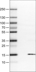
- Experimental details
- Lane 1: Marker [kDa]Lane 2: Negative control (vector only transfected HEK293T lysate) Lane 3: S100A4 Over-expression Lysate (Co-expressed with a C-terminal myc-DDK tag (~3.1 kDa) in mammalian HEK293T cells, LY418991)

Supportive validation
- Submitted by
- Atlas Antibodies (provider)
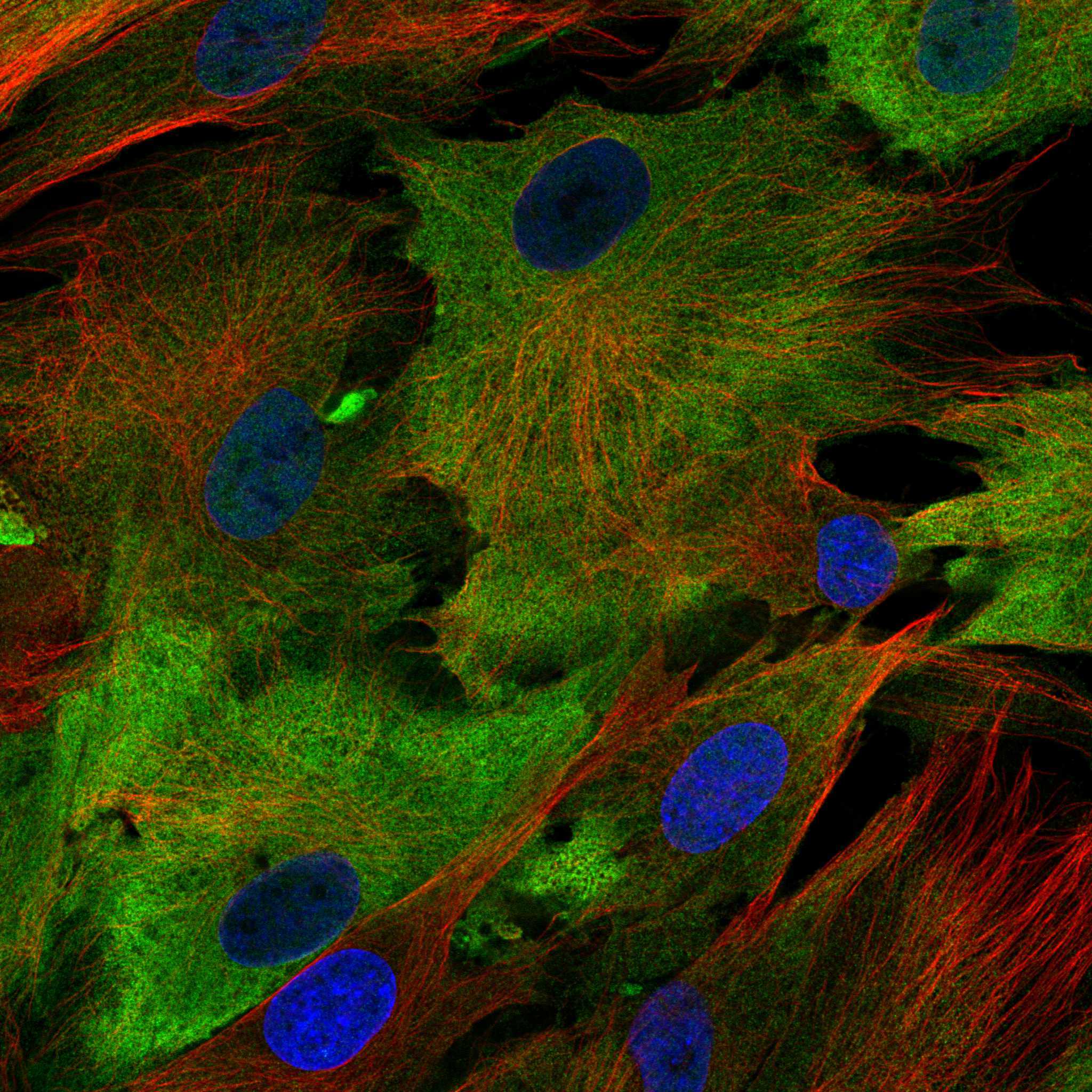
- Main image

- Experimental details
- Immunofluorescence staining of BJ cells using the anti-S100A4 monoclonal antibody, showing specific staining in the plasma membrane in green. Microtubule- and nuclear probes are visualized in red and blue, respectively (where available).
- Sample type
- HUMAN
Enhanced validation
Supportive validation
- Submitted by
- Atlas Antibodies (provider)
- Enhanced method
- Orthogonal validation
- Main image
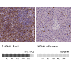
- Experimental details
- Immunohistochemistry analysis in human tonsil and pancreas tissues using AMAb90598 antibody. Corresponding S100A4 RNA-seq data are presented for the same tissues.
- Sample type
- HUMAN

Supportive validation
- Submitted by
- Atlas Antibodies (provider)
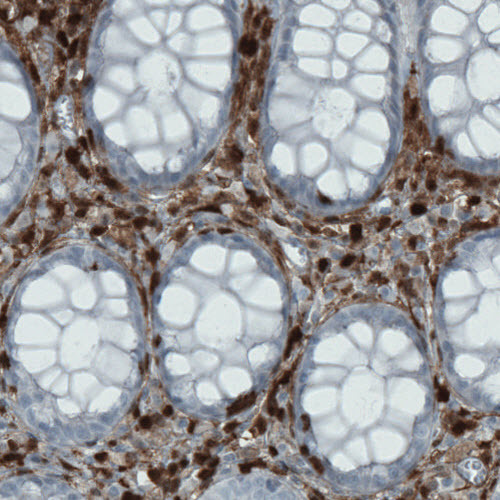
- Main image

- Experimental details
- Immunohistochemical staining of human rectum shows strong immunoreactivity in a subset of lymphoid cells.
- Submitted by
- Atlas Antibodies (provider)
- Main image

- Experimental details
- Immunohistochemical staining of human stomach shows strong positivty in lymphoid cells.

- Submitted by
- Atlas Antibodies (provider)
- Main image
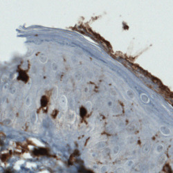
- Experimental details
- Immunohistochemical staining of human skin shows strong immunoreactivity in Langerhans cells.

- Submitted by
- Atlas Antibodies (provider)
- Main image
- Experimental details
- Immunohistochemical staining of human uterus shows positivity in lymphoid cells but not in uterine glands or stroma cells.

- Submitted by
- Atlas Antibodies (provider)
- Main image

- Experimental details
- Immunohistochemical staining of human tonsil shows strong cytoplasmic positivity, mainly in non - germinal center cells.

- Submitted by
- Atlas Antibodies (provider)
- Main image

- Experimental details
- Immunohistochemical staining of human rectum shows strong cytoplasmic positivity in lymphoid cells.

- Submitted by
- Atlas Antibodies (provider)
- Main image

- Experimental details
- Immunohistochemical staining of human endometrium shows strong cytoplasmic positivity in lymphoid cells.

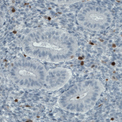
- Submitted by
- Atlas Antibodies (provider)
- Main image

- Experimental details
- Immunohistochemical staining of human pancreas shows no positivity in either islets of Langerhans or exocrine glandular cells as expected.
