 Explore
 Validate
 Learn
LearnH00006275-M01
antibody from Abnova Corporation
Targeting: S100A4
18A2, 42A, CAPL, FSP1, MTS1, P9KA, PEL98
 Western blot
Antibody data
- Antibody Data
- Antigen structure
- References [5]
- Comments [0]
- Validations
- Western blot [3]
- ELISA [1]
- Immunocytochemistry [1]
Submit
Validation data
Reference
Comment
Report error
- Product number
- H00006275-M01 - Provider product page

- Provider
- Abnova Corporation
- Proper citation
- Abnova Corporation Cat#H00006275-M01, RRID:AB_425664
- Product name
- S100A4 monoclonal antibody (M01), clone 1F12-1G7
- Antibody type
- Monoclonal
- Description
- Mouse monoclonal antibody raised against a full length recombinant S100A4.
- Antigen sequence
MACPLEKALDVMVSTFHKYSGKEGDKFKLNKSELK
ELLTRELPSFLGKRTDEAAFQKLMSNLDSNRDNEV
DFQEYCVFLSCIAMMCDEFFEGFPDKQPRKK- Isotype
- IgG
- Antibody clone number
- 1F12-1G7
- Storage
- Store at -20°C or lower. Aliquot to avoid repeated freezing and thawing.
Submitted references Cancer-initiating cells from colorectal cancer patients escape from T cell-mediated immunosurveillance in vitro through membrane-bound IL-4.
MiR-21/Smad 7 signaling determines TGF-β1-induced CAF formation.
Androgen receptor expression in satellite cells of the neonatal levator ani of the rat.
Identification of novel molecular markers through transcriptomic analysis in human fetal and adult corneal endothelial cells.
Potential role for S100A4 in the disruption of the blood-brain barrier in collagen-induced arthritic mice, an animal model of rheumatoid arthritis.
Volonté A, Di Tomaso T, Spinelli M, Todaro M, Sanvito F, Albarello L, Bissolati M, Ghirardelli L, Orsenigo E, Ferrone S, Doglioni C, Stassi G, Dellabona P, Staudacher C, Parmiani G, Maccalli C
Journal of immunology (Baltimore, Md. : 1950) 2014 Jan 1;192(1):523-32
Journal of immunology (Baltimore, Md. : 1950) 2014 Jan 1;192(1):523-32
MiR-21/Smad 7 signaling determines TGF-β1-induced CAF formation.
Li Q, Zhang D, Wang Y, Sun P, Hou X, Larner J, Xiong W, Mi J
Scientific reports 2013;3:2038
Scientific reports 2013;3:2038
Androgen receptor expression in satellite cells of the neonatal levator ani of the rat.
Swift-Gallant A, Monks DA
Developmental neurobiology 2013 Jun;73(6):448-54
Developmental neurobiology 2013 Jun;73(6):448-54
Identification of novel molecular markers through transcriptomic analysis in human fetal and adult corneal endothelial cells.
Chen Y, Huang K, Nakatsu MN, Xue Z, Deng SX, Fan G
Human molecular genetics 2013 Apr 1;22(7):1271-9
Human molecular genetics 2013 Apr 1;22(7):1271-9
Potential role for S100A4 in the disruption of the blood-brain barrier in collagen-induced arthritic mice, an animal model of rheumatoid arthritis.
Nishioku T, Furusho K, Tomita A, Ohishi H, Dohgu S, Shuto H, Yamauchi A, Kataoka Y
Neuroscience 2011 Aug 25;189:286-92
Neuroscience 2011 Aug 25;189:286-92
No comments: Submit comment
Supportive validation
- Submitted by
- Abnova Corporation (provider)
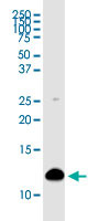
- Main image

- Experimental details
- S100A4 monoclonal antibody (M01), clone 1F12-1G7 Western Blot analysis of S100A4 expression in Hela ( Cat # L013V1 ).

- Submitted by
- Abnova Corporation (provider)
- Main image

- Experimental details
- Western Blot analysis of S100A4 expression in transfected 293T cell line by S100A4 monoclonal antibody (M01), clone 1F12-1G7.Lane 1: S100A4 transfected lysate(11.7 KDa).Lane 2: Non-transfected lysate.

- Submitted by
- Abnova Corporation (provider)
- Main image

- Experimental details
- S100A4 monoclonal antibody (M01), clone 1F12-1G7. Western Blot analysis of S100A4 expression in Raw 264.7.
Supportive validation
- Submitted by
- Abnova Corporation (provider)
- Main image

- Experimental details
- Detection limit for recombinant GST tagged S100A4 is approximately 0.1ng/ml as a capture antibody.
- Validation comment
- Sandwich ELISA (Recombinant protein)
- Protocol
- Protocol

Supportive validation
- Submitted by
- Abnova Corporation (provider)
- Main image

- Experimental details
- Immunofluorescence of monoclonal antibody to S100A4 on HeLa cell. [antibody concentration 15 ug/ml]
- Validation comment
- Immunofluorescence
- Protocol
- Protocol
