MA1-219
antibody from Invitrogen Antibodies
Targeting: PROM1
AC133, CD133, CORD12, MCDR2, PROML1, RP41, STGD4
Antibody data
- Antibody Data
- Antigen structure
- References [1]
- Comments [0]
- Validations
- Western blot [2]
- Immunocytochemistry [1]
- Immunohistochemistry [1]
Submit
Validation data
Reference
Comment
Report error
- Product number
- MA1-219 - Provider product page

- Provider
- Invitrogen Antibodies
- Product name
- Prom1 Monoclonal Antibody (2F8C5)
- Antibody type
- Monoclonal
- Antigen
- Purifed from natural sources
- Description
- MA1-219 Prom1 antibody detects Prom1 (CD133) by western blot and immunofluorescence. MA1-219 Prom1 antibody detects Prom1 by western blot at ~130 kDa.
- Reactivity
- Human
- Host
- Mouse
- Isotype
- IgG
- Antibody clone number
- 2F8C5
- Vial size
- 100 µg
- Concentration
- 1 mg/mL
- Storage
- -20°C
Submitted references Planar polarity in primate cone photoreceptors: a potential role in Stiles Crawford effect phototropism.
Verschueren A, Boucherit L, Ferrari U, Fouquet S, Nouvel-Jaillard C, Paques M, Picaud S, Sahel JA
Communications biology 2022 Jan 24;5(1):89
Communications biology 2022 Jan 24;5(1):89
No comments: Submit comment
Supportive validation
- Submitted by
- Invitrogen Antibodies (provider)
- Main image

- Experimental details
- Knockout of Prom1 was achieved by CRISPR-Cas9 genome editing using LentiArray™ Lentiviral sgRNA (Product # A32042, Assay ID CRISPR924819_LV) and LentiArray Cas9 Lentivirus (Product # A32064). Western blot analysis of Prom1 was performed by loading 30 µg of HT-29 Cas9 (Lane 1) and HT-29 Prom1 KO (Lane 2) whole cell extracts. The samples were electrophoresed using NuPAGE™ Novex™ 4-12% Bis-Tris Protein Gel (Product # NP0322BOX). Resolved proteins were then transferred onto a nitrocellulose membrane (Product # IB23001) by iBlot® 2 Dry Blotting System (Product # IB21001). The blot was probed with Prom1 Monoclonal Antibody (2F8C5) (Product # MA1-219, 1:1000 dilution) and Goat anti-Mouse IgG (H+L) Superclonal™ Recombinant Secondary Antibody, HRP (Product # A28177, 1:5000 dilution) using the iBright™ FL 1500 (Product #A44115). Chemiluminescent detection was performed using SuperSignal™ West Dura Extended Duration Substrate (Product # 34076). Loss of signal upon CRISPR mediated knockout (KO) using the LentiArray™ CRISPR product line confirms that antibody is specific to Prom1.
- Submitted by
- Invitrogen Antibodies (provider)
- Main image

- Experimental details
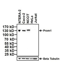
- Western blot analysis of Prom1 was performed by loading 20 µg of indicated whole cell lysates and 7 µL of PageRuler Prestained Protein Ladder (Product # 26616) per well onto a 4-20% Tris-Glycine polyacrylamide gel (Product # WT4202BX10). Proteins were transferred to a nitrocellulose membrane using the G2 Blotter (Product # 62288), and blocked with 5% Milk in TBST for 1 hour at room temperature. Prom1 was detected at 130 kDa using a Prom1 mouse monoclonal antibody (Product # MA1-219, upper panel) at a dilution of 1:1000 in blocking buffer overnight at 4°C on a rocking platform, followed by a Goat anti-mouse IgG (H+L) Secondary Antibody, HRP conjugate (Product # 31430) at a dilution of 1:20000 for at least 30 minutes at room temperature. Beta-Tubulin was detected using mouse monoclonal antibody (Product # MA5-16308, lower panel), followed by a Goat anti-mouse IgG (H+L) Secondary Antibody, HRP conjugate (Product # 31430) at a dilution of 1:20000 for at least 30 minutes at room temperature. Chemiluminescent detection was performed using SuperSignal West Dura (Product # 34076).
Supportive validation
- Submitted by
- Invitrogen Antibodies (provider)
- Main image

- Experimental details
- Immunofluorescent analysis of Prom1 (green) in HT-29 and HeLa cells. The cells were fixed with 4% paraformaldehyde for 15 minutes at -20c, permeabilized with 0.1% Triton X-100 for 15 minutes, and blocked with 3% BSA for 30 minutes at room temperature. Cells were stained with a Prom1 mouse monoclonal antibody (Product # MA1-219) at a dilution of 1:100 in blocking buffer overnight at 4°C, and then incubated with a Goat anti-Mouse IgG (H+L) Secondary Antibody, Alexa Fluor Plus 488 conjugate (Product # A32731) at a dilution of 1:1000 for at least 30 minutes at a room temperature in the dark (green). F-actin (red) was stained with Dylight 554 Phalloidin. Nuclei (blue) were stained with Hoechst 33342 (Product # 62249). Images were taken on a Thermo Scientific ToxInsight Instrument at 20X magnification.
Supportive validation
- Submitted by
- Invitrogen Antibodies (provider)
- Main image

- Experimental details
- Immunohistochemistry was performed on human cervix and human cerebellum. Tissue was deparaffinized with xylene, followed by rehydration in sequential washes of 100% ethanol, 95% ethanol, 80% ethanol, and water. To expose target proteins, antigen retrieval was performed using 10mM sodium citrate (pH 6.0) and heated for 20 min in Lab Vision PT Module (Product # A80400012). Following antigen retrieval, tissues were blocked in a 10% goat serum (Product # 50062Z) in wash buffer solution for 30 minutes at room temperature and endogenous peroxidase activity quenched with Peroxidase Suppressor (Product # 35000). Tissue was then probed with a Prom1 mouse monoclonal antibody (Product # MA1-219) at a dilution of 1:50 in 10% goat serum in wash buffer for 1 hour at room temperature in a humidified chamber. Negative control cervix tissue received no primary antibody. Tissues were washed extensively with PBST, and detection was performed using a SuperBoost™ goat anti-mouse Poly HRP secondary antibody reagent (Product # B40961) followed by colorimetric detection using DAB Quanto (Product # TA-125-QHDX). Tissues were then counterstained with hematoxylin (Product # TA-125-MH), mounted and imaged on EVOS FL Auto Imaging Station.
 Explore
 Validate
 Learn
Learn Western blot