 Explore
 Validate
 Learn
Learn Western blot
 Immunohistochemistry
Antibody data
- Antibody Data
- Antigen structure
- References [31]
- Comments [0]
- Validations
- Immunohistochemistry [1]
- Other assay [13]
Submit
Validation data
Reference
Comment
Report error
- Product number
- MA1-813 - Provider product page

- Provider
- Invitrogen Antibodies
- Product name
- CRALBP Monoclonal Antibody (B2)
- Antibody type
- Monoclonal
- Antigen
- Recombinant full-length protein
- Description
- MA1-813 detects CRALBP protein in bovine, human, mouse, non-human primate and rat samples. MA1-813 has successfully been used in Western blot, immunohistochemistry, immunoprecipitation, and immunofluorescence procedures. By Western blot, this antibody detects a 37 kDa protein representing human recombinant CRALBP. The MA1-813 immunogen is purified human recombinant CRALBP.
- Reactivity
- Human, Mouse, Rat, Bovine
- Host
- Mouse
- Isotype
- IgG
- Antibody clone number
- B2
- Vial size
- 100 µg
- Concentration
- 1 mg/mL
- Storage
- -20° C, Avoid Freeze/Thaw Cycles
Submitted references Derivation and Characterization of Murine and Amphibian Müller Glia Cell Lines.
A Novel Method of Mouse RPE Explant Culture and Effective Introduction of Transgenes Using Adenoviral Transduction for In Vitro Studies in AMD.
Human MiniPromoters for ocular-rAAV expression in ON bipolar, cone, corneal, endothelial, Müller glial, and PAX6 cells.
Glial cells of the human fovea.
CNTF Prevents Development of Outer Retinal Neovascularization Through Upregulation of CxCl10.
Rescue of the MERTK phagocytic defect in a human iPSC disease model using translational read-through inducing drugs.
Directing Differentiation of Pluripotent Stem Cells Toward Retinal Pigment Epithelium Lineage.
Systemic Injection of RPE65-Programmed Bone Marrow-Derived Cells Prevents Progression of Chronic Retinal Degeneration.
Targeting the cAMP and Transforming Growth Factor-β Pathway Increases Proliferation to Promote Re-Epithelialization of Human Stem Cell-Derived Retinal Pigment Epithelium.
Microphthalmia-associated transcription factor regulates the visual cycle genes Rlbp1 and Rdh5 in the retinal pigment epithelium.
Features specific to retinal pigment epithelium cells derived from three-dimensional human embryonic stem cell cultures - a new donor for cell therapy.
AAV-mediated RLBP1 gene therapy improves the rate of dark adaptation in Rlbp1 knockout mice.
A FOXM1 Dependent Mesenchymal-Epithelial Transition in Retinal Pigment Epithelium Cells.
SOCS3 in retinal neurons and glial cells suppresses VEGF signaling to prevent pathological neovascular growth.
Assessment of retinal function and morphology in aging Ccl2 knockout mice.
Retinoid uptake, processing, and secretion in human iPS-RPE support the visual cycle.
Loss of Müller's cells and photoreceptors in macular telangiectasia type 2.
Highly sensitive in vitro methods for detection of residual undifferentiated cells in retinal pigment epithelial cells derived from human iPS cells.
GABA-mediated induction of early neuronal markers expression in postnatal rat progenitor cells in culture.
Induction of differentiation by pyruvate and DMEM in the human retinal pigment epithelium cell line ARPE-19.
Diabetes-induced peroxynitrite impairs the balance of pro-nerve growth factor and nerve growth factor, and causes neurovascular injury.
Ghrelin modulates physiologic and pathologic retinal angiogenesis through GHSR-1a.
Genetic deletion of COX-2 diminishes VEGF production in mouse retinal Müller cells.
Perifoveal müller cell depletion in a case of macular telangiectasia type 2.
Neuroprotection in the juvenile rat model of light-induced retinopathy: evidence suggesting a role for FGF-2 and CNTF.
Stage-specific association of apolipoprotein A-I and E in developing mouse retina.
Cellular retinaldehyde-binding protein interacts with ERM-binding phosphoprotein 50 in retinal pigment epithelium.
Temporal/spatial expression of retinoid binding proteins and RAR isoforms in the postnatal lung.
Direct channeling of retinoic acid between cellular retinoic acid-binding protein II and retinoic acid receptor sensitizes mammary carcinoma cells to retinoic acid-induced growth arrest.
Topological and epitope mapping of the cellular retinaldehyde-binding protein from retina.
Immunocytochemical localization of two retinoid-binding proteins in vertebrate retina.
Gallo RA, Qureshi F, Strong TA, Lang SH, Pino KA, Dvoriantchikova G, Pelaez D
Translational vision science & technology 2022 Apr 1;11(4):4
Translational vision science & technology 2022 Apr 1;11(4):4
A Novel Method of Mouse RPE Explant Culture and Effective Introduction of Transgenes Using Adenoviral Transduction for In Vitro Studies in AMD.
Shang P, Stepicheva NA, Liu H, Chowdhury O, Franks J, Sun M, Hose S, Ghosh S, Yazdankhah M, Strizhakova A, Stolz DB, Zigler JS Jr, Sinha D
International journal of molecular sciences 2021 Nov 5;22(21)
International journal of molecular sciences 2021 Nov 5;22(21)
Human MiniPromoters for ocular-rAAV expression in ON bipolar, cone, corneal, endothelial, Müller glial, and PAX6 cells.
Korecki AJ, Cueva-Vargas JL, Fornes O, Agostinone J, Farkas RA, Hickmott JW, Lam SL, Mathelier A, Zhou M, Wasserman WW, Di Polo A, Simpson EM
Gene therapy 2021 Jun;28(6):351-372
Gene therapy 2021 Jun;28(6):351-372
Glial cells of the human fovea.
Delaunay K, Khamsy L, Kowalczuk L, Moulin A, Nicolas M, Zografos L, Lassiaz P, Behar-Cohen F
Molecular vision 2020;26:235-245
Molecular vision 2020;26:235-245
CNTF Prevents Development of Outer Retinal Neovascularization Through Upregulation of CxCl10.
Bucher F, Aguilar E, Marra KV, Rapp J, Arnold J, Diaz-Aguilar S, Lange C, Agostini H, Schlunck G, Stahl A, Friedlander M
Investigative ophthalmology & visual science 2020 Aug 3;61(10):20
Investigative ophthalmology & visual science 2020 Aug 3;61(10):20
Rescue of the MERTK phagocytic defect in a human iPSC disease model using translational read-through inducing drugs.
Ramsden CM, Nommiste B, R Lane A, Carr AF, Powner MB, J K Smart M, Chen LL, Muthiah MN, Webster AR, Moore AT, Cheetham ME, da Cruz L, Coffey PJ
Scientific reports 2017 Mar 3;7(1):51
Scientific reports 2017 Mar 3;7(1):51
Directing Differentiation of Pluripotent Stem Cells Toward Retinal Pigment Epithelium Lineage.
Choudhary P, Booth H, Gutteridge A, Surmacz B, Louca I, Steer J, Kerby J, Whiting PJ
Stem cells translational medicine 2017 Feb;6(2):490-501
Stem cells translational medicine 2017 Feb;6(2):490-501
Systemic Injection of RPE65-Programmed Bone Marrow-Derived Cells Prevents Progression of Chronic Retinal Degeneration.
Qi X, Pay SL, Yan Y, Thomas J Jr, Lewin AS, Chang LJ, Grant MB, Boulton ME
Molecular therapy : the journal of the American Society of Gene Therapy 2017 Apr 5;25(4):917-927
Molecular therapy : the journal of the American Society of Gene Therapy 2017 Apr 5;25(4):917-927
Targeting the cAMP and Transforming Growth Factor-β Pathway Increases Proliferation to Promote Re-Epithelialization of Human Stem Cell-Derived Retinal Pigment Epithelium.
Choudhary P, Gutteridge A, Impey E, Storer RI, Owen RM, Whiting PJ, Bictash M, Benn CL
Stem cells translational medicine 2016 Jul;5(7):925-37
Stem cells translational medicine 2016 Jul;5(7):925-37
Microphthalmia-associated transcription factor regulates the visual cycle genes Rlbp1 and Rdh5 in the retinal pigment epithelium.
Wen B, Li S, Li H, Chen Y, Ma X, Wang J, Lu F, Qu J, Hou L
Scientific reports 2016 Feb 15;6:21208
Scientific reports 2016 Feb 15;6:21208
Features specific to retinal pigment epithelium cells derived from three-dimensional human embryonic stem cell cultures - a new donor for cell therapy.
Wu W, Zeng Y, Li Z, Li Q, Xu H, Yin ZQ
Oncotarget 2016 Apr 19;7(16):22819-33
Oncotarget 2016 Apr 19;7(16):22819-33
AAV-mediated RLBP1 gene therapy improves the rate of dark adaptation in Rlbp1 knockout mice.
Choi VW, Bigelow CE, McGee TL, Gujar AN, Li H, Hanks SM, Vrouvlianis J, Maker M, Leehy B, Zhang Y, Aranda J, Bounoutas G, Demirs JT, Yang J, Ornberg R, Wang Y, Martin W, Stout KR, Argentieri G, Grosenstein P, Diaz D, Turner O, Jaffee BD, Police SR, Dryja TP
Molecular therapy. Methods & clinical development 2015;2:15022
Molecular therapy. Methods & clinical development 2015;2:15022
A FOXM1 Dependent Mesenchymal-Epithelial Transition in Retinal Pigment Epithelium Cells.
Choudhary P, Dodsworth BT, Sidders B, Gutteridge A, Michaelides C, Duckworth JK, Whiting PJ, Benn CL
PloS one 2015;10(6):e0130379
PloS one 2015;10(6):e0130379
SOCS3 in retinal neurons and glial cells suppresses VEGF signaling to prevent pathological neovascular growth.
Sun Y, Ju M, Lin Z, Fredrick TW, Evans LP, Tian KT, Saba NJ, Morss PC, Pu WT, Chen J, Stahl A, Joyal JS, Smith LE
Science signaling 2015 Sep 22;8(395):ra94
Science signaling 2015 Sep 22;8(395):ra94
Assessment of retinal function and morphology in aging Ccl2 knockout mice.
Vessey KA, Waugh M, Jobling AI, Phipps JA, Ho T, Trogrlic L, Greferath U, Fletcher EL
Investigative ophthalmology & visual science 2015 Jan 27;56(2):1238-52
Investigative ophthalmology & visual science 2015 Jan 27;56(2):1238-52
Retinoid uptake, processing, and secretion in human iPS-RPE support the visual cycle.
Muñiz A, Greene WA, Plamper ML, Choi JH, Johnson AJ, Tsin AT, Wang HC
Investigative ophthalmology & visual science 2014 Jan 9;55(1):198-209
Investigative ophthalmology & visual science 2014 Jan 9;55(1):198-209
Loss of Müller's cells and photoreceptors in macular telangiectasia type 2.
Powner MB, Gillies MC, Zhu M, Vevis K, Hunyor AP, Fruttiger M
Ophthalmology 2013 Nov;120(11):2344-52
Ophthalmology 2013 Nov;120(11):2344-52
Highly sensitive in vitro methods for detection of residual undifferentiated cells in retinal pigment epithelial cells derived from human iPS cells.
Kuroda T, Yasuda S, Kusakawa S, Hirata N, Kanda Y, Suzuki K, Takahashi M, Nishikawa S, Kawamata S, Sato Y
PloS one 2012;7(5):e37342
PloS one 2012;7(5):e37342
GABA-mediated induction of early neuronal markers expression in postnatal rat progenitor cells in culture.
Ramírez M, Hernández-Montoya J, Sánchez-Serrano SL, Ordaz B, Ferraro S, Quintero H, Peña-Ortega F, Lamas M
Neuroscience 2012 Nov 8;224:210-22
Neuroscience 2012 Nov 8;224:210-22
Induction of differentiation by pyruvate and DMEM in the human retinal pigment epithelium cell line ARPE-19.
Ahmado A, Carr AJ, Vugler AA, Semo M, Gias C, Lawrence JM, Chen LL, Chen FK, Turowski P, da Cruz L, Coffey PJ
Investigative ophthalmology & visual science 2011 Sep 9;52(10):7148-59
Investigative ophthalmology & visual science 2011 Sep 9;52(10):7148-59
Diabetes-induced peroxynitrite impairs the balance of pro-nerve growth factor and nerve growth factor, and causes neurovascular injury.
Ali TK, Al-Gayyar MM, Matragoon S, Pillai BA, Abdelsaid MA, Nussbaum JJ, El-Remessy AB
Diabetologia 2011 Mar;54(3):657-68
Diabetologia 2011 Mar;54(3):657-68
Ghrelin modulates physiologic and pathologic retinal angiogenesis through GHSR-1a.
Zaniolo K, Sapieha P, Shao Z, Stahl A, Zhu T, Tremblay S, Picard E, Madaan A, Blais M, Lachapelle P, Mancini J, Hardy P, Smith LE, Ong H, Chemtob S
Investigative ophthalmology & visual science 2011 Jul 23;52(8):5376-86
Investigative ophthalmology & visual science 2011 Jul 23;52(8):5376-86
Genetic deletion of COX-2 diminishes VEGF production in mouse retinal Müller cells.
Yanni SE, McCollum GW, Penn JS
Experimental eye research 2010 Jul;91(1):34-41
Experimental eye research 2010 Jul;91(1):34-41
Perifoveal müller cell depletion in a case of macular telangiectasia type 2.
Powner MB, Gillies MC, Tretiach M, Scott A, Guymer RH, Hageman GS, Fruttiger M
Ophthalmology 2010 Dec;117(12):2407-16
Ophthalmology 2010 Dec;117(12):2407-16
Neuroprotection in the juvenile rat model of light-induced retinopathy: evidence suggesting a role for FGF-2 and CNTF.
Joly S, Pernet V, Chemtob S, Di Polo A, Lachapelle P
Investigative ophthalmology & visual science 2007 May;48(5):2311-20
Investigative ophthalmology & visual science 2007 May;48(5):2311-20
Stage-specific association of apolipoprotein A-I and E in developing mouse retina.
Kurumada S, Onishi A, Imai H, Ishii K, Kobayashi T, Sato SB
Investigative ophthalmology & visual science 2007 Apr;48(4):1815-23
Investigative ophthalmology & visual science 2007 Apr;48(4):1815-23
Cellular retinaldehyde-binding protein interacts with ERM-binding phosphoprotein 50 in retinal pigment epithelium.
Nawrot M, West K, Huang J, Possin DE, Bretscher A, Crabb JW, Saari JC
Investigative ophthalmology & visual science 2004 Feb;45(2):393-401
Investigative ophthalmology & visual science 2004 Feb;45(2):393-401
Temporal/spatial expression of retinoid binding proteins and RAR isoforms in the postnatal lung.
Hind M, Corcoran J, Maden M
American journal of physiology. Lung cellular and molecular physiology 2002 Mar;282(3):L468-76
American journal of physiology. Lung cellular and molecular physiology 2002 Mar;282(3):L468-76
Direct channeling of retinoic acid between cellular retinoic acid-binding protein II and retinoic acid receptor sensitizes mammary carcinoma cells to retinoic acid-induced growth arrest.
Budhu AS, Noy N
Molecular and cellular biology 2002 Apr;22(8):2632-41
Molecular and cellular biology 2002 Apr;22(8):2632-41
Topological and epitope mapping of the cellular retinaldehyde-binding protein from retina.
Crabb JW, Gaur VP, Garwin GG, Marx SV, Chapline C, Johnson CM, Saari JC
The Journal of biological chemistry 1991 Sep 5;266(25):16674-83
The Journal of biological chemistry 1991 Sep 5;266(25):16674-83
Immunocytochemical localization of two retinoid-binding proteins in vertebrate retina.
Bunt-Milam AH, Saari JC
The Journal of cell biology 1983 Sep;97(3):703-12
The Journal of cell biology 1983 Sep;97(3):703-12
No comments: Submit comment
Supportive validation
- Submitted by
- Invitrogen Antibodies (provider)
- Main image
- Experimental details
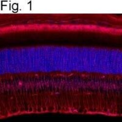
- Immunostaining of CRALBP in frozen mouse retina sections using Product # MA1-813.

Supportive validation
- Submitted by
- Invitrogen Antibodies (provider)
- Main image

- Experimental details
- NULL

- Submitted by
- Invitrogen Antibodies (provider)
- Main image

- Experimental details
- NULL

- Submitted by
- Invitrogen Antibodies (provider)
- Main image

- Experimental details
- Fig 1 A Mesenchymal-Epithelial Transition with temporal FOXM1 expression during RPE culture. A. Immunocytochemistry was performed for CRALBP, ZO1, alphaSMA and Ki67 at Day 0 (D0), Day 3 (D3) and Day 35 (D35). Arrowheads point towards Ki67 positive nuclei. B. Microarray heatmap of the expression profiles of the top 250 genes, ranked by the significance of their expression changes, over time in culture. Raw expression data are mean centred and scaled to unit variance prior to clustering. A schematic of the scaled expression is shown on the right where individual gene profiles are in light grey and the mean expression profile is shown in black. C. Microarray heatmap showing transcript expression for a panel of representative markers over a timecourse of RPE culture. D. Immunocytochemistry for FOXM1 at Day 2 and Day 14 of RPE culture. Arrowheads point towards FOXM1 positive nuclei. E. Quantification of immunocytochemistry showing percentage of nuclei staining positive for FOXM1 over time. Bars represent Mean +- SD (n = 3). F. Expression of FOXM1 transcript measured using qPCR (relative to housekeeping genes ACTB and GAPDH ) in iPSC derived RPE, human foetal RPE and ARPE19 cells over time. Bars represent Mean +- SD (n = 3)

- Submitted by
- Invitrogen Antibodies (provider)
- Main image

- Experimental details
- Figure 1 Patterns of eGFP expression mediated by reporter vectors in mouse and monkey retinas. ( a ) Following dissection of the neural retina from a mouse eye injected with scAAV8p RLBP1 (short)-eGFP, the remaining eye cup was fixed, flat-mounted and imaged. The photograph shows the large geographic regions of RPE that express eGFP. ( b-p ) Neural retina was fixed, sectioned and costained with cell-specific antibody markers. ( b-d ) Images of a retina transduced by the scAAV8-p RLBP1 (short)-eGFP vector showing eGFP, CRALBP, and the merger of eGFP and CRALBP signals demonstrate eGFP expression in CRALBP-expressing Muller cells spanning the retina from the external limiting membrane to the end feet in the inner limiting membrane; ( e-h ) eGFP, vimentin, eGFP+vimentin merged, and eGFP+GFAP merged indicate that eGFP is coexpressed with vimentin and GFAP in Muller cell processes of the inner nuclear layer and the inner limiting membrane where vimentin and GFAP are in intermediate filaments of Muller cells. ( i-k ) Images of eGFP, blue opsin, and eGFP+blue opsin merged reveal that eGFP does not colocalize with cone photoreceptors that express blue opsin, and ( m-o ) images of eGFP, green opsin, and eGFP+green opsin merged show that eGFP does not colocalize with cone photoreceptors that express green opsin. As examples of off-target photoreceptor expression, images from a ( l ) merger of images of eGFP+blue opsin expression from a retina injected with ssAAV8-p BEST -eGFP injected

- Submitted by
- Invitrogen Antibodies (provider)
- Main image

- Experimental details
- Fig 6 BMP/Wnt signalling is required for MET. A. Immunocytochemistry for PMEL17 where cells are seeded at either low density (16000 cells/cm 2 ) in the presence or absence of BMP4/7 (top left) or at high density (25000 cells/cm 2 ) in the presence or absence of Wnt5B (bottom left) and cultured for a period of 14 days. Also shown is the expression of BEST1 under the same conditions (top and bottom right). ACTB and B2M are used as housekeeping genes. Bars represent Mean + SD (n = 3). B. Quantification of immunocytochemistry for % CRALBP at Day 21 where cells are either treated with media alone (Control) or media supplemented with 10muM LDN-193189 added at Day 2,4,6,8,11,14 or 18. * indicates significant difference between control and compound treatment (One way ANOVA with Dunnett's multiple comparisons). C. Quantification of immunocytochemistry for % CRALBP at Day 28 where cells are either treated with media alone (Control) or media supplemented with 10muM WAY-262611 added at Day 2,7,14 or 21. * indicates significant difference between control and compound treatment (One way ANOVA with Dunnett's multiple comparisons). D. qPCR based measurement of BMP7 and Wnt5B transcript expression at Day 10 post siFOXM1 transfection (relative to transfection with non-targeting siRNA used as a control). GAPDH , HPRT1 and IPO8 were used as housekeeping genes. Bars represent Mean + SD (n = 3). P

- Submitted by
- Invitrogen Antibodies (provider)
- Main image

- Experimental details
- Figure 2 MITF is required and sufficient to induce RLBP1 and RDH5 expression in ARPE-19 cells. ( A ) The expression of RLBP1 and RDH5 in ARPE-19 cells transfected with scramble siRNA (si-C) or various concentrations of MITF siRNA (si-MITF). Increased si-MITF concentrations resulted in enhanced MITF knockdown. The expression of the MITF target gene TYR was reduced concomitantly with the reduction of MITF . With the reduction in MITF expression, the expression of RLBP1 and RDH5 was also decreased. ( B ) Protein expression of RLBP1 and RDH5 was decreased when MITF was knocked-down in ARPE-19 cells. TUBULIN was used as an internal protein loading control. ( C ) The RNA and protein levels of RLBP1 and RDH5 in EGFP- and MITF- overexpressing ARPE-19. Both RNA and protein expression of RLBP1 and RDH5 were increased when MITF was overexpressed in ARPE19 cells compared to overexpression of EGFP . The experiments were performed in triplicate and the error bars represent the S.E. *indicates P < 0.05.

- Submitted by
- Invitrogen Antibodies (provider)
- Main image

- Experimental details
- Figure 4 The differentiation of 3D-RPE cells ( A-C ) RT-qPCR analysis of expression of CRALBP, BEST1 and RPE65 in 3D-RPE cells versus time-matched SD-RPE cells at day 40, day 60 and day 100 after differentiation respectively (bars: mean + s.d.; n = 3). All samples were normalized to glyceraldehyde 3-phosphate dehydrogenase (GAPDH). Single asterisk (*) indicated p value < 0.05 and double asterisk (**) indicated p value < 0.01. ( D-F ) Immunostaining of CRALBP and BEST1 of 3D-RPE and SD-RPE cells at day 40, day 60 and day 100 after differentiation respectively. Z-stack fluorescent images with cross-section side views were displayed to show BEST1 location.

- Submitted by
- Invitrogen Antibodies (provider)
- Main image

- Experimental details
- Figure 3 Characterization of the Rlbp1 -/- mouse. ( a ) The horizontal bar is a schematic representation of the Rlbp 1 targeted deletion in the Lexicon TF0133 mouse line. Primers used for genotyping are depicted as arrows (P-9, P-3, and P-14). ( b ) PCR analysis of DNA from Rlbp1 -/- , Rlbp1 +/- , and Rlbp1 +/+ mice using primers P-9, P-3, and P-14 show the expected 854 bp amplicon of the WT allele and the 358 bp amplicon of the allele carrying the deletion. ( c,d ) Paraffin sections of Rlbp1 +/+ ( c ) and Rlbp1 -/- ( d ) mouse eyes stained with an antibody against CRALBP. ( e,f ) Hematoxylin and eosin stained paraffin sections of 16-month-old Rlbp1 +/+ ( e ) and Rlbp1 -/- ( f ) mouse eyes. ( g ) Lysates of neural retina from uninjected Rlbp1 -/- (KO uninj.), Rlbp1 -/- injected with 1 x 10 9 vg/eye of scAAV8-p RLBP1 (short)-h RLBP1 (KO, vector inj.), and uninjected Rlbp1 +/+ (WT uninj.) mice were analyzed by western blot using a primary antibody against CRALBP. Arrow, end feet of Muller cells; arrow head, Muller cell processes; diamond, cell bodies of inner nuclear layer; asterisk, RPE; scale bars, 50 um in c+d and 100 um in e+f .

- Submitted by
- Invitrogen Antibodies (provider)
- Main image

- Experimental details
- Fig. 6 Development of NR2E1 MiniPromoters for Muller glia cells. A Bioinformatic design of first-generation Ple264, cutdown to produce second-generation Ple315, and redesign to produce second-generation Ple316. ENCODE segments that potentially regulate the expression of the NR2E1 gene are highlighted horizontally. Vertically highlighted genomic regions correspond to their color-matched segments included in the MiniPromoter designs and are numbered as promoter(s) (P) and regulatory region(s) (RR). B Postnatal day 4 intravenous injection of Ple316-EmGFP, harvested 4 weeks later, led to robust expression with cell bodies in the inner nuclear layer (INL) and processes that span the retina. Scale bar, 100 um. (For ubiquitous smCBA promoter see Fig. S1A .) C Adult intravitreal injection of Ple316-EmGFP, harvested 4 weeks later, was quantified for epifluorescence intensity in CRALBP-positive cells, and showed no significant difference when compared to smCBA. (For smCBA see Fig. S1D ). D Adult intravitreal injection of Ple316-EmGFP, harvested 4 weeks later, led to robust expression in the Muller glia cells, as indicated by co-staining with Muller glia cell marker CRALBP. Scale bar, 20 um. (For smCBA see Fig. S1D .) EmGFP emerald green fluorescent protein, GCL ganglion cell layer, IPL inner plexiform layer, N number of cells counted, ONL outer nuclear layer, OPL outer plexiform layer, TF transcription factor, TSS transcription start site. Green, anti-GFP; blue, Hoechst; red, anti-CRAL

- Submitted by
- Invitrogen Antibodies (provider)
- Main image

- Experimental details
- Figure 4 Coimmunostaining of P3 foveal glial cells with the Muller cell marker CRALBP, GFAP, and vimentin. Coimmunostaining of foveal cryosections from P3 with cellular retinaldehyde-binding protein (CRALBP), which is a specific marker of Muller cells in humans, glial fibrillar acidic protein (GFAP), and vimentin (VIM), which stains all glial origin cells, shows that the GFAP-positive cells at the roof of the foveal pit ( A , B , C , D , and inset) are stained with vimentin ( G , H , and inset) but do not express CRALBP ( E , F , and inset). I : Costaining with GFAP and vimentin shows that the GFAP-positive cells at the roof of the pit (arrow) are faintly stained with vimentin and are distinct from the vimentin-positive Muller glial cell extensions (red arrow). GCL, ganglion cell layer; INL, inner nuclear layer; ONL, outer nuclear layer; HL, Henle's fiber layer; OLM, outer limiting membrane.

- Submitted by
- Invitrogen Antibodies (provider)
- Main image

- Experimental details
- Figure 3 Visual cycle proteins were abundantly expressed in the RPE flatmounts during the first 3 days in culture, but gradually decreased over time. RPE flatmounts obtained from 2- and 9-month-old wild-type C57BL/6J mice were cultured in complete medium for up to 7 days. RPE flatmounts treated with H 2 O 2 were cultured for 16 h. ( A ) Protein lysates were prepared from the RPE flatmounts and subjected to western blotting analysis to detect the levels of visual cycle proteins RPE65 and CRALBP. Statistical analysis was performed using one-way ANOVA. * p < 0.05, ** p < 0.01, *** p < 0.001, **** p < 0.0001. n = 3. ( B ) RPE65 and CRALBP protein levels were also measured by immunofluorescence. Data from both the western blotting and immunofluorescence showed the gradual reduction in the two proteins in RPE flatmounts over time. Scale bar = 20 mum.

- Submitted by
- Invitrogen Antibodies (provider)
- Main image

- Experimental details
- Figure 1. Immunophenotypic characterization of mouse MG cell line RG17. (A) Phase contrast images and immunofluorescence staining of primary MG cells and passage 20 RG17 cells. RG17 demonstrate typical MG morphology in culture and express many well-characterized MG markers. Scale bar, 50 um and applies to all immunofluorescence images. (B) Expression of characteristic MG genes in primary MG and RG17 cells (passages 20-25). The geometric means and geometric standard deviations ( N = 3 independent cultures) are graphed. An unpaired t -test was applied with a P value of

- Submitted by
- Invitrogen Antibodies (provider)
- Main image

- Experimental details
- Figure 2. Immunophenotypic characterization of X laevis MG cell line XG69. (A) Phase contrast images and immunofluorescence staining of primary MG and XG69 cell line at passage 20. (B) Expression of MG genes in primary MG and XG69 cells (passages 20-25). The geometric means and geometric standard deviations ( N = 3 independent cultures) are graphed. An unpaired t -test was applied with a P value of
