 Explore
 Validate
 Learn
Learn Western blot
Antibody data
- Antibody Data
- Antigen structure
- References [5]
- Comments [0]
- Validations
- Western blot [1]
- Immunocytochemistry [1]
- Immunohistochemistry [6]
- Other assay [2]
Submit
Validation data
Reference
Comment
Report error
- Product number
- PA5-95137 - Provider product page

- Provider
- Invitrogen Antibodies
- Product name
- Collagen I Polyclonal Antibody
- Antibody type
- Polyclonal
- Antigen
- Synthetic peptide
- Description
- Reconstitute with 0.2 mL of distilled water to yield a concentration of 500 µg/mL.
- Reactivity
- Human, Mouse, Rat
- Host
- Rabbit
- Isotype
- IgG
- Vial size
- 100 µg
- Concentration
- 500 µg/mL
- Storage
- Store at 4°C short term. For long term storage, store at -20°C, avoiding freeze/thaw cycles.
Submitted references Paclitaxel Induces Epidermal Molecular Changes and Produces Subclinical Alterations in the Skin of Gynecological Cancer Patients.
Leflunomide Induces Dose-Dependent Lung Injury in Mice via Stimulating Vimentin and NLRP3 Inflammasome Production.
LTBP4 affects renal fibrosis by influencing angiogenesis and altering mitochondrial structure.
Additively manufactured BaTiO(3) composite scaffolds: A novel strategy for load bearing bone tissue engineering applications.
mRNA and long non-coding RNA expression profiling of human periodontal ligament cells under tension loading.
Montero P, Pérez-Leal M, Pérez-Fidalgo JA, Sanz C, Estornut C, Roger I, Milara J, Cervantes A, Cortijo J
Cancers 2022 Feb 23;14(5)
Cancers 2022 Feb 23;14(5)
Leflunomide Induces Dose-Dependent Lung Injury in Mice via Stimulating Vimentin and NLRP3 Inflammasome Production.
El-Sherbiny M, Atef H, Eladl MA, Mohamed AS, El-Shafey M, Ali HS, Zaitone SA, Alomar SY, Alqahtani SAM, Aloyouni SY, Attia MA
Frontiers in pharmacology 2021;12:631216
Frontiers in pharmacology 2021;12:631216
LTBP4 affects renal fibrosis by influencing angiogenesis and altering mitochondrial structure.
Su CT, Jao TM, Urban Z, Huang YJ, See DHW, Tsai YC, Lin WC, Huang JW
Cell death & disease 2021 Oct 13;12(10):943
Cell death & disease 2021 Oct 13;12(10):943
Additively manufactured BaTiO(3) composite scaffolds: A novel strategy for load bearing bone tissue engineering applications.
Mancuso E, Shah L, Jindal S, Serenelli C, Tsikriteas ZM, Khanbareh H, Tirella A
Materials science & engineering. C, Materials for biological applications 2021 Jul;126:112192
Materials science & engineering. C, Materials for biological applications 2021 Jul;126:112192
mRNA and long non-coding RNA expression profiling of human periodontal ligament cells under tension loading.
Lin Y, Cheng T, Zhu S, Gu M, Jin L, Yang Y
European journal of orthodontics 2021 Dec 1;43(6):698-707
European journal of orthodontics 2021 Dec 1;43(6):698-707
No comments: Submit comment
Supportive validation
- Submitted by
- Invitrogen Antibodies (provider)
- Main image

- Experimental details
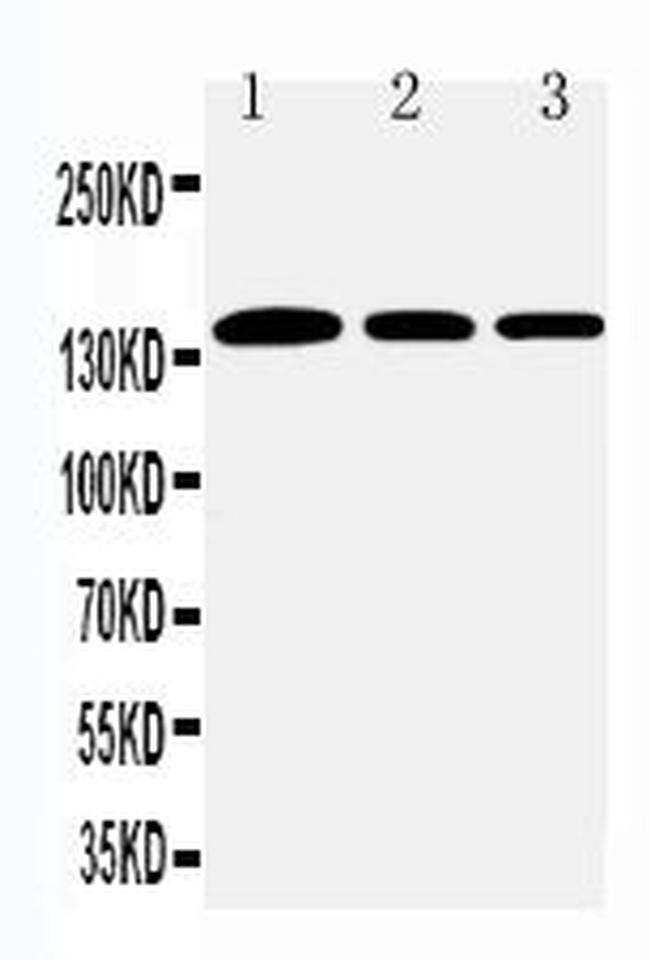
- Western blot analysis of Collagen I in Lane 1: rat lung tissue lysate Lane 2: human placenta tissue lysate Lane 3: rat testis tissue lysate After Electrophoresis, proteins were transferred to a Nitrocellulose membrane at 150mA for 50-90 minutes. Electrophoresis was performed with 5-20% SDS-PAGE gel (70V, Stacking gel; 90V Resolving gel, Time: 2-3 hours), transferred to a nitrocellulose membrane and blocked using 5% Non-fat Milk/TBS (1.5 hrs at room temperature). Samples were incubated with Collagen I polyclonal antibody (Product # PA5-95137) using a 0.5 µg/mL dilution, followed by a goat anti-rabbit IgG-HRP at a dilution of 1:10,000, and developed with enhanced chemiluminescence (ECL).
Supportive validation
- Submitted by
- Invitrogen Antibodies (provider)
- Main image

- Experimental details
- Immunofluorescence analysis of Collagen I in NIH3T3 cell. Antigen retrieval was performed on the tissue using citrate buffer (pH 6, 20 min) and blocked with 10% goat serum. Samples were incubated with Collagen I polyclonal antibody (Product # PA5-95137) at a 1 µg/mL dilution, followed by biotinylated goat anti-rabbit IgG (30 min, 37°C) using a dilution and developed with Strepavidin-Biotin-Complex/DAB.
Supportive validation
- Submitted by
- Invitrogen Antibodies (provider)
- Main image

- Experimental details
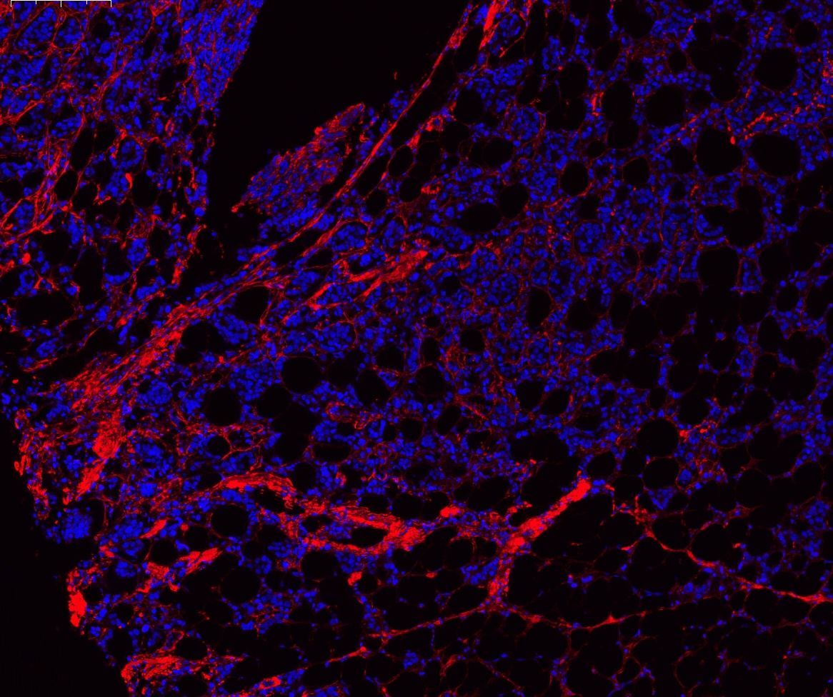
- Immunofluorescence analysis of Collagen I in human tonsil tissues. Antigen retrieval was performed on the tissue using citrate buffer (pH 6, 20 min) and blocked with 10% goat serum. Samples were incubated with Collagen I polyclonal antibody (Product # PA5-95137) at a 1 µg/mL dilution, followed by Cy3 conjugated Goat anti-rabbit IgG using a 1:100 dilution and DAPI.
- Submitted by
- Invitrogen Antibodies (provider)
- Main image

- Experimental details
- Immunofluorescence analysis of Collagen I in human tonsil tissues. Antigen retrieval was performed on the tissue using citrate buffer (pH 6, 20 min) and blocked with 10% goat serum. Samples were incubated with Collagen I polyclonal antibody (Product # PA5-95137) at a 1 µg/mL dilution, followed by Cy3 conjugated Goat anti-rabbit IgG using a 1:100 dilution and DAPI.

- Submitted by
- Invitrogen Antibodies (provider)
- Main image

- Experimental details
- Immunofluorescence analysis of Collagen I in human mammary tissue tissues. Antigen retrieval was performed on the tissue using citrate buffer (pH 6, 20 min) and blocked with 10% goat serum. Samples were incubated with Collagen I polyclonal antibody (Product # PA5-95137) at a 1 µg/mL dilution, followed by Cy3 conjugated Goat anti-rabbit IgG using a 1:100 dilution and DAPI.

- Submitted by
- Invitrogen Antibodies (provider)
- Main image

- Experimental details
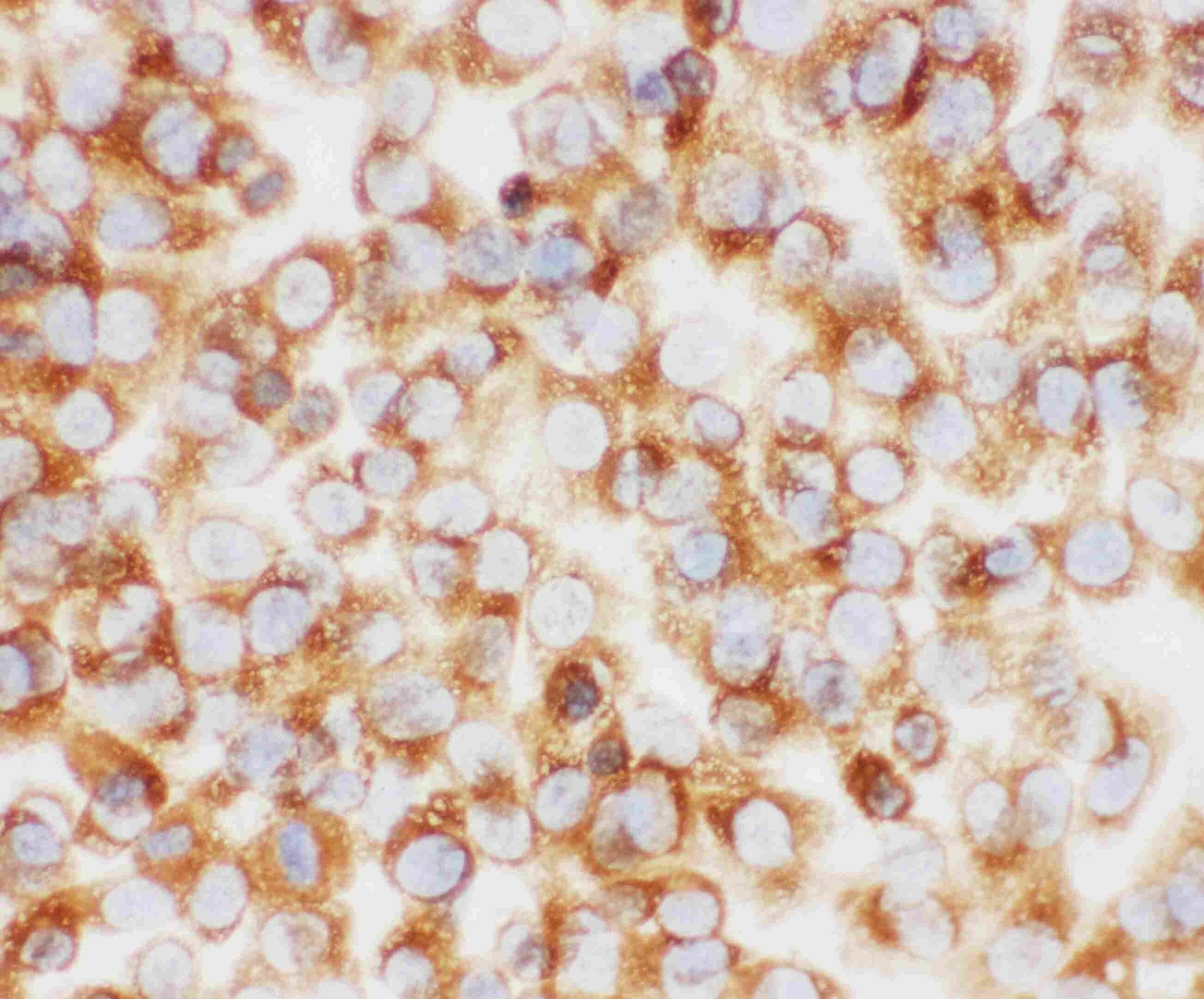
- Immunohistochemistry analysis of Collagen I in paraffin-embedded human placenta tissue. Antigen retrieval was performed on the tissue using citrate buffer (pH 6, 20 min) and blocked with 10% goat serum. Samples were incubated with Collagen I polyclonal antibody (Product # PA5-95137) at a 1 µg/mL dilution, followed by biotinylated goat anti-rabbit IgG (30 min, 37°C), and developed with Strepavidin-Biotin-Complex and DAB.

- Submitted by
- Invitrogen Antibodies (provider)
- Main image

- Experimental details
- Immunohistochemistry analysis of Collagen I in paraffin-embedded rat lung tissues. Antigen retrieval was performed on the tissue using citrate buffer (pH 6, 20 min) and blocked with 10% goat serum. Samples were incubated with Collagen I polyclonal antibody (Product # PA5-95137) at a 1 µg/mL dilution, followed by biotinylated goat anti-rabbit IgG (30 min, 37°C), and developed with Strepavidin-Biotin-Complex and DAB.

- Submitted by
- Invitrogen Antibodies (provider)
- Main image

- Experimental details
- Immunohistochemistry analysis of Collagen I in paraffin-embedded human intestinal cancer tissues. Antigen retrieval was performed on the tissue using citrate buffer (pH 6, 20 min) and blocked with 10% goat serum. Samples were incubated with Collagen I polyclonal antibody (Product # PA5-95137) at a 1 µg/mL dilution, followed by biotinylated goat anti-rabbit IgG (30 min, 37°C), and developed with Strepavidin-Biotin-Complex and DAB.

Supportive validation
- Submitted by
- Invitrogen Antibodies (provider)
- Main image
- Experimental details
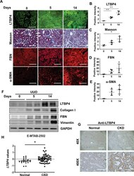
- Fig. 1 LTBP4 expression is associated with tubulointerstitial fibrosis in mouse and human kidneys. A - F Wild-type mice were subjected to tubulointerstitial fibrosis by unilateral ureteral obstruction (UUO) for the indicated time points. A Representative histological images with Masson's trichrome and immunofluorescence staining of kidneys after UUO. Scale bars, 100 mum. Computer-assisted quantitative analyses of histological images for Ltbp4 ( B ), Masson's trichrome (Masson) ( C ), Fibronectin (FBN) ( D ), and alpha-SMA ( E ) are shown. F Protein expression of Ltbp4, collagen I, fibronectin, and vimentin in the kidneys of mice subjected to UUO, as detected by immunoblotting. GAPDH served as an internal control. Representative images from three independent experiments are shown above. n = 2 mice in 0-day group; n = 4 mice in 5- and 14-day groups. G Representative immunohistochemical images showing that LTBP4 was predominantly expressed in the kidneys of a patient with diabetic nephropathy compared to a normal control individual. Scale bars: 1 mm in x40 and 100 mum in x400 magnifications. Representative images from three independent experiments are shown above. ( H ) LTBP4 was upregulated in fibrotic kidneys, which was analysed in microarray data set: ArrayExpress_E-MTAB-2502. Normal: health individual; CKD: chronic kidney disease. Data are presented as the mean +- SEM. * p < 0.05, ** p < 0.01.

- Submitted by
- Invitrogen Antibodies (provider)
- Main image

- Experimental details
- Figure 1. Tensile loading enhanced the osteogenic differentiation of human periodontal ligament (PDL) cells. (A) Quantitative polymerase chain reaction (qPCR) analysis of the relative mRNA expression of collagen type I ( COL1 ) and runt-related transcription factor 2 ( RUNX2 ) in PDL cells subjected or not to tensile loading for 12 hours. The results are normalized to the expression of TATA-box-binding protein ( TBP ) and peptidylprolyl isomerase B ( PPIB ) and expressed as means +- standard deviations. (B) Western blot analysis and relative quantification of COL1 and RUNX2 proteins in PDL cells subjected or not to tensile loading for 3 days. (C) Visualization and relative quantification of Alizarin Red S staining (ARS) in PDL cells subjected or not to tensile loading. * P < 0.05, ** P < 0.01.
