Explore
 Validate
 Learn
Learn Western blot
Antibody data
- Antibody Data
- Antigen structure
- References [0]
- Comments [0]
- Validations
- Western blot [1]
- Immunocytochemistry [1]
- Immunohistochemistry [10]
Submit
Validation data
Reference
Comment
Report error
- Product number
- AMAb90664 - Provider product page

- Provider
- Atlas Antibodies
- Proper citation
- Atlas Antibodies Cat#AMAb90664, RRID:AB_2665625
- Product name
- Anti-CTCF
- Antibody type
- Monoclonal
- Reactivity
- Human
- Host
- Mouse
- Conjugate
- Unconjugated
- Antigen sequence
QNQTDGGEVVQDVNSSVQMVMMEQLDPTLLQMKTE
VMEGTVAPEAEAAVDDTQIITLQVVNMEEQPINIG
ELQLVQVPVPVTVPVATTSVEELQGAYENEVSKEG
LAESEPMICHTLPLPEGFQVVKVGANGEVETLEQG
ELPPQEDP- Epitope
- Binds to an epitope located within the peptide sequence NMEEQPINIGELQLV as determined by overlapping synthetic peptides.
- Isotype
- IgG
- Antibody clone number
- CL0305
- Vial size
- 100 µl
- Storage
- Store at +4°C for short term storage. Long time storage is recommended at -20°C.
No comments: Submit comment
Supportive validation
- Submitted by
- Atlas Antibodies (provider)
- Main image

- Experimental details
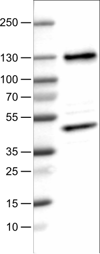
- Lane 1: Marker [kDa]Lane 2: Human cell line U-251 MG
Supportive validation
- Submitted by
- Atlas Antibodies (provider)
- Main image
- Experimental details
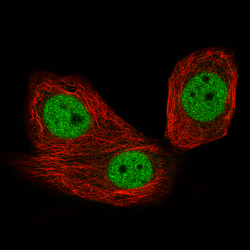
- Immunofluorescence staining of MCF7 cells using the Anti-CTCF monoclonal antibody, showing specific staining in the nucleoplasm in green. Microtubule- and nuclear probes are visualized in red and blue, respectively (where available).
- Sample type
- HUMAN

Supportive validation
- Submitted by
- Atlas Antibodies (provider)
- Main image
- Experimental details
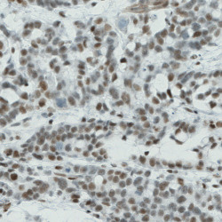
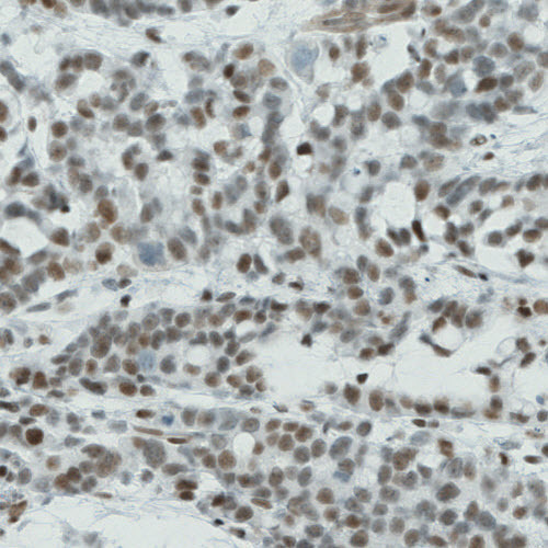
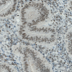
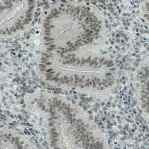
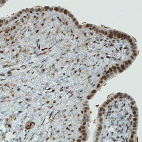
- Immunohistochemical staining of human stomach cancer shows nuclear immunoreactivity in tumor cells.
- Submitted by
- Atlas Antibodies (provider)
- Main image
- Experimental details
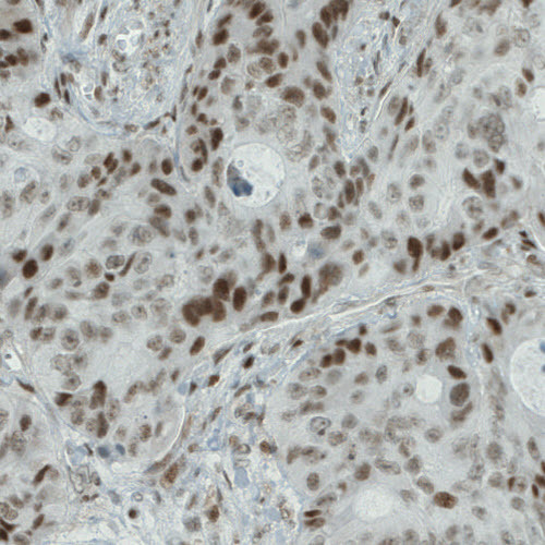
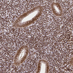
- Immunohistochemical staining of human colorectal cancer shows nuclear positivity in tumor cells.
- Submitted by
- Atlas Antibodies (provider)
- Main image
- Experimental details
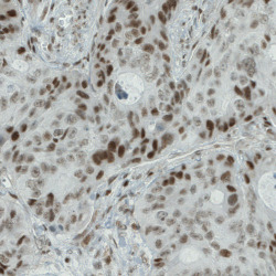
- Immunohistochemical staining of human prostate cancer shows nuclear positivity in tumor cells.

- Submitted by
- Atlas Antibodies (provider)
- Main image
- Experimental details
- Immunohistochemical staining of human stomach shows nuclear immunoreactivity in glandular cells.
- Submitted by
- Atlas Antibodies (provider)
- Main image
- Experimental details
- Immunohistochemical staining of human uterus shows nuclear positivity in glandular and stromal cells.
- Submitted by
- Atlas Antibodies (provider)
- Main image

- Experimental details
- Immuohistochemical staining of human fallopian tube shows nuclear staining in epithelial cells.
- Submitted by
- Atlas Antibodies (provider)
- Main image
- Experimental details
- Immunohistochemical staining of human endometrium shows moderate nuclear positivity in glandular and stromal cells.

- Submitted by
- Atlas Antibodies (provider)
- Main image

- Experimental details
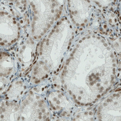
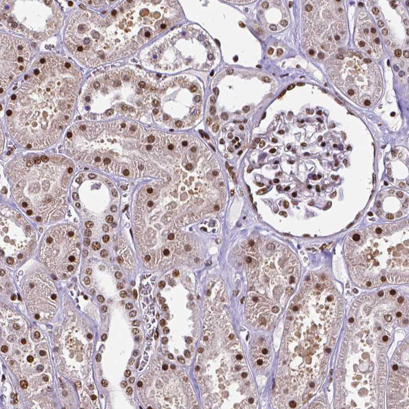
- Immunohistochemical staining of human kidney shows moderate to strong nuclear positivity in cells in tubuli and glomeruli.
- Submitted by
- Atlas Antibodies (provider)
- Main image

- Experimental details
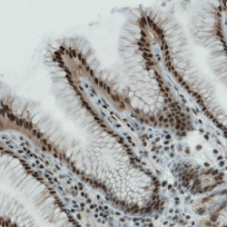
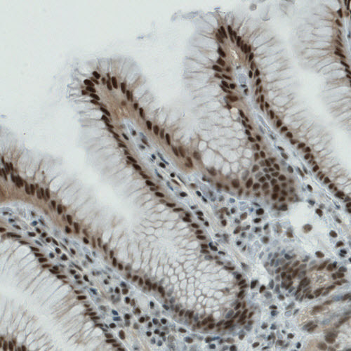
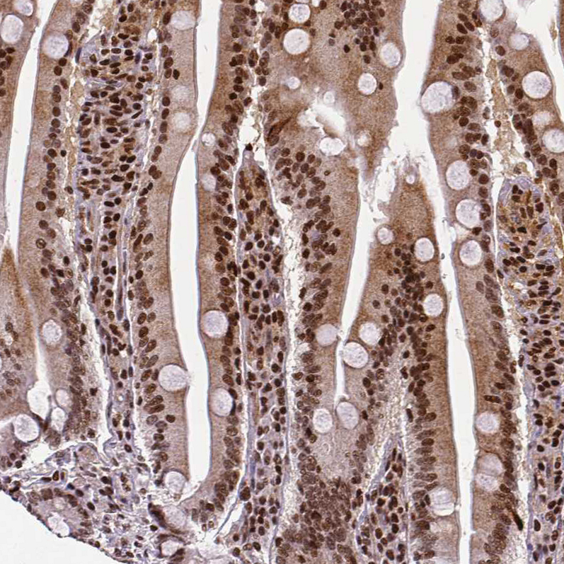
- Immunohistochemical staining of human small intestine shows strong nuclear positivity in glandular cells.
- Submitted by
- Atlas Antibodies (provider)
- Main image

- Experimental details
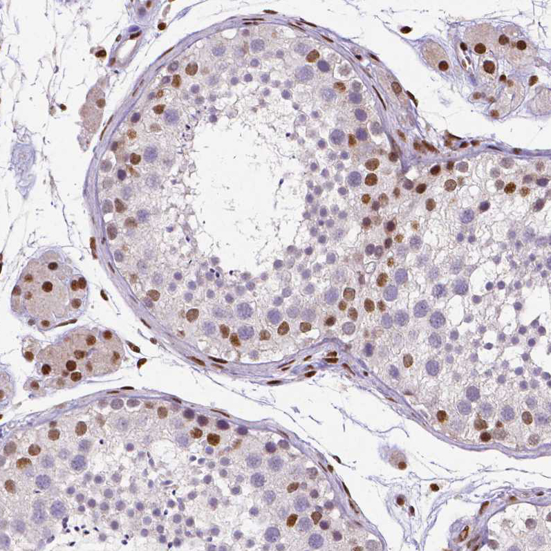
- Immunohistochemical staining of human testis shows moderate nuclear positivity in a subset of cells in seminiferous ducts.