 Explore
 Validate
 Learn
Learn Western blot
 Immunohistochemistry
Antibody data
- Antibody Data
- Antigen structure
- References [4]
- Comments [0]
- Validations
- Immunohistochemistry [7]
Submit
Validation data
Reference
Comment
Report error
- Product number
- HPA012012 - Provider product page

- Provider
- Atlas Antibodies
- Proper citation
- Atlas Antibodies Cat#HPA012012, RRID:AB_1855371
- Product name
- Anti-PIGR
- Antibody type
- Polyclonal
- Reactivity
- Human
- Host
- Rabbit
- Conjugate
- Unconjugated
- Antigen sequence
LLFSVVINQLRLSDAGQYLCQAGDDSNSNKKNADL
QVLKPEPELVYEDLRGSVTFHCALGPEVANVAKFL
CRQSSGENCDVVVNTLGKRAPAFEGRILLNPQDKD
GSFSVVITGLRKEDAGRYLCGAHS- Isotype
- IgG
- Vial size
- 100 µl
- Storage
- Store at +4°C for short term storage. Long time storage is recommended at -20°C.
Submitted references Reduced expression of the polymeric immunoglobulin receptor in pancreatic and periampullary adenocarcinoma signifies tumour progression and poor prognosis.
Expression and prognostic significance of the polymeric immunoglobulin receptor in epithelial ovarian cancer.
Expression and prognostic significance of the polymeric immunoglobulin receptor in esophageal and gastric adenocarcinoma.
Age-related expression of the polymeric immunoglobulin receptor (pIgR) in the gastric mucosa of young pigs.
Fristedt R, Elebro J, Gaber A, Jonsson L, Heby M, Yudina Y, Nodin B, Uhlén M, Eberhard J, Jirström K
PloS one 2014;9(11):e112728
PloS one 2014;9(11):e112728
Expression and prognostic significance of the polymeric immunoglobulin receptor in epithelial ovarian cancer.
Berntsson J, Lundgren S, Nodin B, Uhlén M, Gaber A, Jirström K
Journal of ovarian research 2014 Feb 26;7:26
Journal of ovarian research 2014 Feb 26;7:26
Expression and prognostic significance of the polymeric immunoglobulin receptor in esophageal and gastric adenocarcinoma.
Fristedt R, Gaber A, Hedner C, Nodin B, Uhlén M, Eberhard J, Jirström K
Journal of translational medicine 2014 Apr 2;12:83
Journal of translational medicine 2014 Apr 2;12:83
Age-related expression of the polymeric immunoglobulin receptor (pIgR) in the gastric mucosa of young pigs.
Trevisi P, Gandolfi G, Priori D, Messori S, Colombo M, Mazzoni M, Lallès JP, Bosi P
PloS one 2013;8(11):e81473
PloS one 2013;8(11):e81473
No comments: Submit comment
Enhanced validation
Supportive validation
- Submitted by
- Atlas Antibodies (provider)
- Enhanced method
- Orthogonal validation
- Main image
- Experimental details
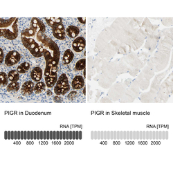
- Immunohistochemistry analysis in human duodenum and skeletal muscle tissues using HPA012012 antibody. Corresponding PIGR RNA-seq data are presented for the same tissues.
- Sample type
- HUMAN

Supportive validation
- Submitted by
- Atlas Antibodies (provider)
- Main image

- Experimental details
- Immunohistochemical staining of human colon shows high expression.
- Sample type
- HUMAN

- Submitted by
- Atlas Antibodies (provider)
- Main image

- Experimental details
- Immunohistochemical staining of human liver shows low expression as expected.
- Sample type
- HUMAN

- Submitted by
- Atlas Antibodies (provider)
- Main image

- Experimental details
- Immunohistochemical staining of human kidney shows moderate cytoplasmic positivity in cells in tubules.
- Sample type
- HUMAN

- Submitted by
- Atlas Antibodies (provider)
- Main image

- Experimental details
- Immunohistochemical staining of human tonsil shows no positivity in non-germinal center cells as expected.
- Sample type
- HUMAN

- Submitted by
- Atlas Antibodies (provider)
- Main image

- Experimental details
- Immunohistochemical staining of human duodenum shows strong cytoplasmic and membranous positivity in glandular cells.
- Sample type
- HUMAN

- Submitted by
- Atlas Antibodies (provider)
- Main image

- Experimental details
- Immunohistochemical staining of human skeletal muscle shows no positivity in myocytes as expected.
- Sample type
- HUMAN
