Explore
 Validate
 Learn
Learn Western blot
 ELISA
ELISAAntibody data
- Antibody Data
- Antigen structure
- References [2]
- Comments [0]
- Validations
- Western blot [1]
- Immunocytochemistry [1]
- Immunohistochemistry [1]
- Flow cytometry [1]
Submit
Validation data
Reference
Comment
Report error
- Product number
- ABIN953145 - Provider product page

- Provider
- antibodies-online
- Product name
- anti-Lecithin-Cholesterol Acyltransferase (LCAT) (AA 292-321), (Middle Region) antibody
- Antibody type
- Polyclonal
- Antigen
- Synthetic peptide - KLH conjugated - corresponding to the central region (between 292-321aa) of human LCAT.
- Description
- Purified through a Protein A column followed by peptide affinity purification
- Reactivity
- Human
- Host
- Rabbit
- Epitope
- AA 292-321,Middle Region
- Vial size
- 0.4 mL
- Concentration
- 0.25 mg/mL
- Storage
- Store undiluted at 2-8°C for one month or (in aliquots) at -20°C for longer.
- Handling
- Avoid repeated freezing and thawing.
Submitted references Investigation of variants identified in caucasian genome-wide association studies for plasma high-density lipoprotein cholesterol and triglycerides levels in Mexican dyslipidemic study samples.
Integrative predictive model of coronary artery calcification in atherosclerosis.
Weissglas-Volkov D, Aguilar-Salinas CA, Sinsheimer JS, Riba L, Huertas-Vazquez A, Ordoñez-Sánchez ML, Rodriguez-Guillen R, Cantor RM, Tusie-Luna T, Pajukanta P
Circulation. Cardiovascular genetics 2010 Feb;3(1):31-8
Circulation. Cardiovascular genetics 2010 Feb;3(1):31-8
Integrative predictive model of coronary artery calcification in atherosclerosis.
McGeachie M, Ramoni RL, Mychaleckyj JC, Furie KL, Dreyfuss JM, Liu Y, Herrington D, Guo X, Lima JA, Post W, Rotter JI, Rich S, Sale M, Ramoni MF
Circulation 2009 Dec 15;120(24):2448-54
Circulation 2009 Dec 15;120(24):2448-54
No comments: Submit comment
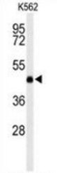
Supportive validation
- Submitted by
- antibodies-online (provider)
- Main image
- Experimental details
- Western blot analysis of LCAT (arrow) in K562 cell line lysates (35ug/lane) using LCAT antibody Cat.-No. AP52450PU-N.

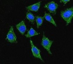
Supportive validation
- Submitted by
- antibodies-online (provider)
- Main image
- Experimental details
- Confocal immunofluorescent analysis with 293 cells using LCAT antibody Cat.-No. AP52450PU-N, followed by Alexa Fluor® 488-conjugated goat anti-rabbit lgG (green). DAPI was used to stain the cell nuclear (blue).

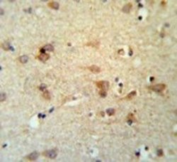
Supportive validation
- Submitted by
- antibodies-online (provider)
- Main image
- Experimental details
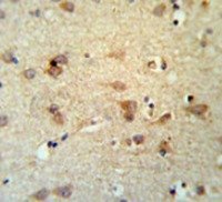
- Immunohistochemistry analysis in brain tissue (Formalin-fixed, Paraffin-embedded) using LCAT antibody Cat.-No. AP52450PU-N, followed by peroxidase conjugation of the secondary antibody and DAB staining. This data demonstrates the use of the LCAT antibody for IHC; Clinical relevance has not been evaluated.
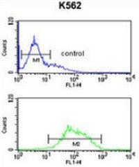
Supportive validation
- Submitted by
- antibodies-online (provider)
- Main image
- Experimental details
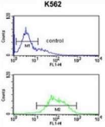
- Flow cytometric analysis of K562 cells (bottom histogram) compared to a negative control cell (top histogram) using LCAT antibody Cat.-No. AP52450PU-N, followed by FITC-conjugated goat-anti-rabbit secondary antibodies.