 Explore
 Validate
 Learn
Learn Western blot
 ELISA
ELISAAntibody data
- Antibody Data
- Antigen structure
- References [3]
- Comments [0]
- Validations
- Western blot [2]
- Immunocytochemistry [2]
- Immunohistochemistry [2]
Submit
Validation data
Reference
Comment
Report error
- Product number
- GTX105765 - Provider product page

- Provider
- GeneTex
- Proper citation
- GeneTex Cat#GTX105765, RRID:AB_11173313
- Product name
- Glutamate Dehydrogenase antibody
- Antibody type
- Polyclonal
- Reactivity
- Human, Mouse, Rat
- Host
- Rabbit
Submitted references Dynamic metabolic patterns tracking neurodegeneration and gliosis following 26S proteasome dysfunction in mouse forebrain neurons.
Structure, kinetic characterization and subcellular localization of the two ribulose 5-phosphate epimerase isoenzymes from Trypanosoma cruzi.
Amniotic fluid proteome analysis from Down syndrome pregnancies for biomarker discovery.
Geiszler PC, Ugun-Klusek A, Lawler K, Pardon MC, Yuchun D, Bai L, Daykin CA, Auer DP, Bedford L
Scientific reports 2018 Mar 19;8(1):4833
Scientific reports 2018 Mar 19;8(1):4833
Structure, kinetic characterization and subcellular localization of the two ribulose 5-phosphate epimerase isoenzymes from Trypanosoma cruzi.
Gonzalez SN, Valsecchi WM, Maugeri D, Delfino JM, Cazzulo JJ
PloS one 2017;12(2):e0172405
PloS one 2017;12(2):e0172405
Amniotic fluid proteome analysis from Down syndrome pregnancies for biomarker discovery.
Cho CK, Smith CR, Diamandis EP
Journal of proteome research 2010 Jul 2;9(7):3574-82
Journal of proteome research 2010 Jul 2;9(7):3574-82
No comments: Submit comment
Supportive validation
- Submitted by
- GeneTex (provider)
- Main image

- Experimental details
- Sample (30 ?g of whole cell lysate) A: HepG2 7.5% SDS PAGE GTX105765 diluted at 1:1000 The HRP-conjugated anti-rabbit IgG antibody (GTX213110-01) was used to detect the primary antibody.

- Submitted by
- GeneTex (provider)
- Main image
- Experimental details
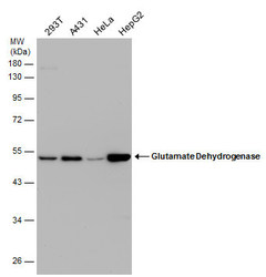
- Various whole cell extracts (30 ?g) were separated by 10% SDS-PAGE, and the membrane was blotted with Glutamate Dehydrogenase antibody (GTX105765) diluted at 1:1000.

Supportive validation
- Submitted by
- GeneTex (provider)
- Main image

- Experimental details
- Glutamate Dehydrogenase antibody detects Glutamate Dehydrogenase protein at cytoplasm by immunofluorescent analysis.Sample: U-87 MG cells were fixed in 4% paraformaldehyde at RT for 15 min.Green: Glutamate Dehydrogenase protein stained by Glutamate Dehydrogenase antibody (GTX105765) diluted at 1:500.Red: beta Tubulin 3/ TUJ1 protein stained by beta Tubulin 3/ TUJ1 antibody (GTX631836) diluted at 1:200.Blue: Hoechst 33342 staining.

- Submitted by
- GeneTex (provider)
- Main image
- Experimental details
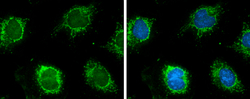
- Glutamate Dehydrogenase antibody detects Glutamate Dehydrogenase protein at mitochondria by immunofluorescent analysis.Sample: HeLa cells were fixed in ice-cold MeOH for 5 min.Green: Glutamate Dehydrogenase protein stained by Glutamate Dehydrogenase antibody (GTX105765) diluted at 1:500.Blue: Hoechst 33342 staining.

Supportive validation
- Submitted by
- GeneTex (provider)
- Main image

- Experimental details
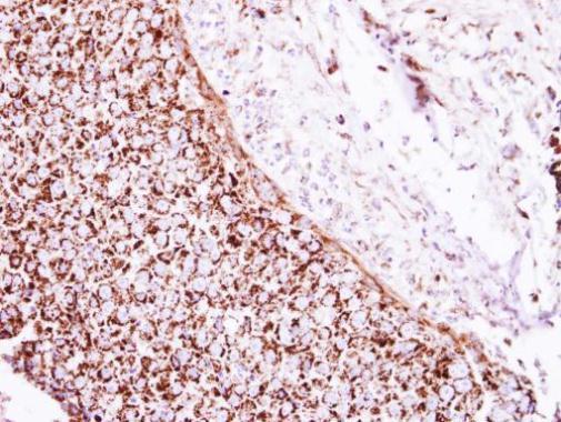
- Immunohistochemical analysis of paraffin-embedded human breast cancer, using Glutamate Dehydrogenase(GTX105765) antibody at 1:250 dilution.
- Submitted by
- GeneTex (provider)
- Main image

- Experimental details
- Glutamate Dehydrogenase antibody detects Glutamate Dehydrogenase protein at cytoplasm in rat brain by immunohistochemical analysis. Sample: Paraffin-embedded rat brain. Glutamate Dehydrogenase antibody (GTX105765) diluted at 1:750.
