 Explore
 Validate
 Learn
Learn Western blot
Antibody data
- Antibody Data
- Antigen structure
- References [9]
- Comments [0]
- Validations
- Western blot [3]
- Immunohistochemistry [1]
Submit
Validation data
Reference
Comment
Report error
- Product number
- AF1197 - Provider product page

- Provider
- R&D Systems
- Product name
- Human/Mouse/Rat Galectin-3 Antibody
- Antibody type
- Polyclonal
- Description
- Antigen Affinity-purified. Detects human, mouse, and rat Galectin-3 in Western blots. Detects mouse Galectin-3 in direct ELISAs. In direct ELISAs, approximately 20% cross-reactivity with recombinant human (rh) Galectin-3 is observed and less than 1% cross-reactivity with recombinant mouse (rm) Galectin-4 and rmGalectin-9 is observed.
- Reactivity
- Human, Mouse, Rat
- Host
- Goat
- Conjugate
- Unconjugated
- Antigen sequence
NP_034835- Isotype
- IgG
- Vial size
- 100 ug
- Concentration
- LYOPH
- Storage
- Use a manual defrost freezer and avoid repeated freeze-thaw cycles. 12 months from date of receipt, -20 to -70 °C as supplied. 1 month, 2 to 8 °C under sterile conditions after reconstitution. 6 months, -20 to -70 °C under sterile conditions after reconstitution.
Submitted references Bush-like integrin filament networks associated with hyaloid vasculature in murine neonate eyes.
Peripheral Inflammation Enhances Microglia Response and Nigral Dopaminergic Cell Death in an in vivo MPTP Model of Parkinson's Disease.
Identifying the Role of Complement in Triggering Neuroinflammation after Traumatic Brain Injury.
Histochemical characteristics of regressing vessels in the hyaloid vascular system of neonatal mice: Novel implication for vascular atrophy.
Acute Complement Inhibition Potentiates Neurorehabilitation and Enhances tPA-Mediated Neuroprotection.
Krüppel-like Factor 3 (KLF3/BKLF) Is Required for Widespread Repression of the Inflammatory Modulator Galectin-3 (Lgals3).
Galectin-1 and galectin-8 have redundant roles in promoting plasma cell formation.
Characterization of RAGE, HMGB1, and S100beta in inflammation-induced preterm birth and fetal tissue injury.
Galectin-3 is an amplifier of inflammation in atherosclerotic plaque progression through macrophage activation and monocyte chemoattraction.
Iwanaga T, Nio-Kobayashi J, Takahashi-Iwanaga H
Biomedical research (Tokyo, Japan) 2019;40(2):79-85
Biomedical research (Tokyo, Japan) 2019;40(2):79-85
Peripheral Inflammation Enhances Microglia Response and Nigral Dopaminergic Cell Death in an in vivo MPTP Model of Parkinson's Disease.
García-Domínguez I, Veselá K, García-Revilla J, Carrillo-Jiménez A, Roca-Ceballos MA, Santiago M, de Pablos RM, Venero JL
Frontiers in cellular neuroscience 2018;12:398
Frontiers in cellular neuroscience 2018;12:398
Identifying the Role of Complement in Triggering Neuroinflammation after Traumatic Brain Injury.
Alawieh A, Langley EF, Weber S, Adkins D, Tomlinson S
The Journal of neuroscience : the official journal of the Society for Neuroscience 2018 Mar 7;38(10):2519-2532
The Journal of neuroscience : the official journal of the Society for Neuroscience 2018 Mar 7;38(10):2519-2532
Histochemical characteristics of regressing vessels in the hyaloid vascular system of neonatal mice: Novel implication for vascular atrophy.
Kishimoto A, Kimura S, Nio-Kobayashi J, Takahashi-Iwanaga H, Park AM, Iwanaga T
Experimental eye research 2018 Jul;172:1-9
Experimental eye research 2018 Jul;172:1-9
Acute Complement Inhibition Potentiates Neurorehabilitation and Enhances tPA-Mediated Neuroprotection.
Alawieh A, Andersen M, Adkins DL, Tomlinson S
The Journal of neuroscience : the official journal of the Society for Neuroscience 2018 Jul 18;38(29):6527-6545
The Journal of neuroscience : the official journal of the Society for Neuroscience 2018 Jul 18;38(29):6527-6545
Krüppel-like Factor 3 (KLF3/BKLF) Is Required for Widespread Repression of the Inflammatory Modulator Galectin-3 (Lgals3).
Knights AJ, Yik JJ, Mat Jusoh H, Norton LJ, Funnell AP, Pearson RC, Bell-Anderson KS, Crossley M, Quinlan KG
The Journal of biological chemistry 2016 Jul 29;291(31):16048-58
The Journal of biological chemistry 2016 Jul 29;291(31):16048-58
Galectin-1 and galectin-8 have redundant roles in promoting plasma cell formation.
Tsai CM, Guan CH, Hsieh HW, Hsu TL, Tu Z, Wu KJ, Lin CH, Lin KI
Journal of immunology (Baltimore, Md. : 1950) 2011 Aug 15;187(4):1643-52
Journal of immunology (Baltimore, Md. : 1950) 2011 Aug 15;187(4):1643-52
Characterization of RAGE, HMGB1, and S100beta in inflammation-induced preterm birth and fetal tissue injury.
Buhimschi CS, Baumbusch MA, Dulay AT, Oliver EA, Lee S, Zhao G, Bhandari V, Ehrenkranz RA, Weiner CP, Madri JA, Buhimschi IA
The American journal of pathology 2009 Sep;175(3):958-75
The American journal of pathology 2009 Sep;175(3):958-75
Galectin-3 is an amplifier of inflammation in atherosclerotic plaque progression through macrophage activation and monocyte chemoattraction.
Papaspyridonos M, McNeill E, de Bono JP, Smith A, Burnand KG, Channon KM, Greaves DR
Arteriosclerosis, thrombosis, and vascular biology 2008 Mar;28(3):433-40
Arteriosclerosis, thrombosis, and vascular biology 2008 Mar;28(3):433-40
No comments: Submit comment
Supportive validation
- Submitted by
- R&D Systems (provider)
- Main image
- Experimental details
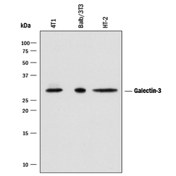
- Detection of Mouse Galectin-3 by Western Blot. Western blot shows lysates of 4T1 mouse breast cancer cell line, Balb/3T3 mouse embryonic fibroblast cell line and HT-2 mouse T cell line. PVDF membrane was probed with 0.05 µg/mL of Goat Anti-Mouse Galectin-3 Antigen Affinity-purified Polyclonal Antibody (Catalog # AF1197) followed by HRP-conjugated Anti-Goat IgG Secondary Antibody (Catalog # HAF017). A specific band was detected for Galectin-3 at approximately 28 kDa (as indicated). This experiment was conducted under reducing conditions and using Immunoblot Buffer Group 1.

- Submitted by
- R&D Systems (provider)
- Main image
- Experimental details
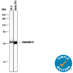
- Detection of Mouse Galectin-3 by Simple WesternTM. Simple Western lane view shows lysates of HT-2 mouse T cell line and Balb/3T3 mouse embryonic fibroblast cell line, loaded at 0.2 mg/mL. A specific band was detected for Galectin-3 at approximately 44 kDa (as indicated) using 10 µg/mL of Goat Anti-Mouse Galectin-3 Antigen Affinity-purified Polyclonal Antibody (Catalog # AF1197) followed by 1:50 dilution of HRP-conjugated Anti-Goat IgG Secondary Antibody (Catalog # HAF109). This experiment was conducted under reducing conditions and using the 12-230 kDa separation system.

- Submitted by
- R&D Systems (provider)
- Main image

- Experimental details
- Detection of Human and Rat Galectin-3 by Western Blot. Western blot shows lysates of MCF-7 human breast cancer cell line, U-118-MG human glioblastoma/astrocytoma cell line, C6 rat glioma cell line, and NR8383 rat alveolar macrophage cell line. PVDF membrane was probed with 0.2 µg/mL of Goat Anti-Mouse Galectin-3 Antigen Affinity-purified Polyclonal Antibody (Catalog # AF1197) followed by HRP-conjugated Anti-Goat IgG Secondary Antibody (Catalog # HAF017). A specific band was detected for Galectin-3 at approximately 28 kDa (as indicated). This experiment was conducted under reducing conditions and using Immunoblot Buffer Group 1.

Supportive validation
- Submitted by
- R&D Systems (provider)
- Main image

- Experimental details
- Galectin-3 in Mouse Embryo. Galectin-3 was detected in immersion fixed frozen sections of mouse embryo (15 d.p.c., cross-section through the eye) using Goat Anti-Human Galectin-3 Antigen Affinity-purified Polyclonal Antibody (Catalog # AF1197 ) at 15 µg/mL overnight at 4 °C. Tissue was stained using the Anti-Goat HRP-DAB Cell & Tissue Staining Kit (brown; Catalog # CTS008) and counterstained with hematoxylin (blue). View our protocol for Chromogenic IHC Staining of Frozen Tissue Sections.
