 Explore
 Validate
 Learn
Learn Western blot
 ELISA
ELISAAntibody data
- Antibody Data
- Antigen structure
- References [2]
- Comments [0]
- Validations
- Western blot [3]
- Immunocytochemistry [3]
- Immunohistochemistry [2]
- Flow cytometry [1]
Submit
Validation data
Reference
Comment
Report error
- Product number
- NB100-59735 - Provider product page

- Provider
- Novus Biologicals
- Proper citation
- Novus Cat#NB100-59735, RRID:AB_892310
- Product name
- Goat Polyclonal xCT Antibody
- Antibody type
- Polyclonal
- Description
- Immunogen affinity purified.
- Reactivity
- Human
- Host
- Goat
- Antigen sequence
KGQTQNFKDAFSGRD- Isotype
- IgG
- Vial size
- 0.1 mg
- Concentration
- 0.5 mg/ml
- Storage
- Store at -20C. Avoid freeze-thaw cycles.
Submitted references Stromal control of cystine metabolism promotes cancer cell survival in chronic lymphocytic leukaemia.
Kaposi's sarcoma-associated herpesvirus fusion-entry receptor: cystine transporter xCT.
Zhang W, Trachootham D, Liu J, Chen G, Pelicano H, Garcia-Prieto C, Lu W, Burger JA, Croce CM, Plunkett W, Keating MJ, Huang P
Nature cell biology 2012 Feb 19;14(3):276-86
Nature cell biology 2012 Feb 19;14(3):276-86
Kaposi's sarcoma-associated herpesvirus fusion-entry receptor: cystine transporter xCT.
Kaleeba JA, Berger EA
Science (New York, N.Y.) 2006 Mar 31;311(5769):1921-4
Science (New York, N.Y.) 2006 Mar 31;311(5769):1921-4
No comments: Submit comment
Supportive validation
- Submitted by
- Novus Biologicals (provider)
- Main image
- Experimental details
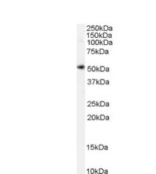
- Western Blot: xCT Antibody [NB100-59735] - Staining of Human Spleen lysate (35 ug protein in RIPA buffer). Detected by chemiluminescence.

- Submitted by
- Novus Biologicals (provider)
- Main image
- Experimental details
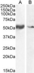
- Western Blot: xCT Antibody [NB100-59735] - Staining of Human Smooth Muscle (A) and Tonsil (B) lysate (35 ug protein in RIPA buffer). Antibody at 0.1 ug/mL. Detected by chemiluminescence.

- Submitted by
- Novus Biologicals (provider)
- Main image
- Experimental details
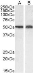
- Western Blot: xCT Antibody [NB100-59735] - Staining of A549 (A) and U2OS (B) cell ysate (35 ug protein in RIPA buffer). Antibody at 0.3 ug/mL. Detected by chemiluminescence.

Supportive validation
- Submitted by
- Novus Biologicals (provider)
- Main image
- Experimental details
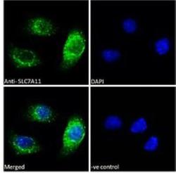
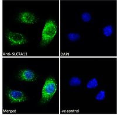
- Immunofluorescence: xCT Antibody [NB100-59735] - Analysis of paraformaldehyde fixed A549 cells, permeabilized with 0.15% Triton. Primary incubation 1hr (10 ug/mL) followed by Alexa Fluor 488 secondary antibody (2 ug/mL), showing vesicle/cytoplasmic staining. The nuclear stain is DAPI (blue). Negative control: Unimmunized goat IgG (10 ug/mL) followed by Alexa Fluor 488 secondary antibody (2 ug/mL).
- Submitted by
- Novus Biologicals (provider)
- Main image

- Experimental details
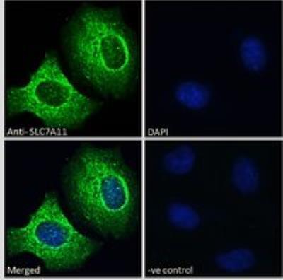
- Immunocytochemistry/Immunofluorescence
- Submitted by
- Novus Biologicals (provider)
- Main image
- Experimental details
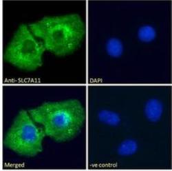
- Immunocytochemistry/Immunofluorescence
Supportive validation
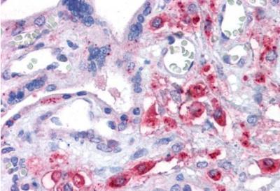
- Submitted by
- Novus Biologicals (provider)
- Main image
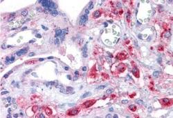
- Experimental details
- Immunohistochemistry-Paraffin: xCT Antibody [NB100-59735] - Staining of Human Placenta. Antibody at 3.75 ug/mL. Steamed antigen retrieval with citrate buffer pH 6, AP-staining.
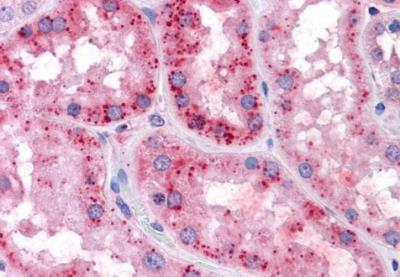
- Submitted by
- Novus Biologicals (provider)
- Main image
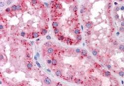
- Experimental details
- Immunohistochemistry-Paraffin: xCT Antibody [NB100-59735] - Staining of Human Kidney. Antibody at 3.75 ug/mL. Steamed antigen retrieval with citrate buffer pH 6, AP-staining.
Supportive validation
- Submitted by
- Novus Biologicals (provider)
- Main image
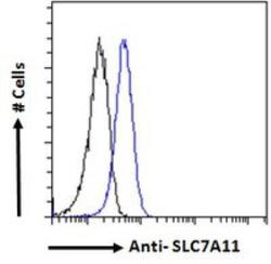
- Experimental details
- Flow Cytometry: xCT Antibody [NB100-59735] - Analysis of paraformaldehyde fixed A549 cells (blue line), permeabilized with 0.5% Triton. Primary incubation 1hr (10 ug/mL) followed by Alexa Fluor 488 secondary antibody (1 ug/mL). IgG control: Unimmunized goat IgG (black line) followed by Alexa Fluor 488 secondary antibody.
