 Explore
 Validate
 Learn
Learn Western blot
 Immunoprecipitation
Antibody data
- Antibody Data
- Antigen structure
- References [0]
- Comments [0]
- Validations
- Western blot [1]
- Immunocytochemistry [1]
Submit
Validation data
Reference
Comment
Report error
- Product number
- NBP2-42199 - Provider product page

- Provider
- Novus Biologicals
- Product name
- Mouse Monoclonal PEX6 Antibody
- Antibody type
- Monoclonal
- Description
- Protein G purified. Less then 50% identity with other PEX proteins.Detects approx 100kDa.
- Reactivity
- Human, Rat
- Host
- Mouse
- Isotype
- IgG
- Vial size
- 0.1 mg
- Concentration
- 1 mg/ml
- Storage
- Store at 4C short term. Aliquot and store at -20C long term. Avoid freeze-thaw cycles.
No comments: Submit comment
Supportive validation
- Submitted by
- Novus Biologicals (provider)
- Main image
- Experimental details
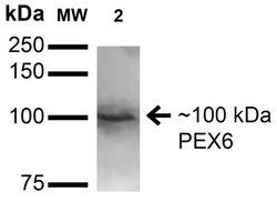
- Western Blot: PEX6 Antibody (S233-8) [NBP2-42199] - analysis of Rat Brain showing detection of 100 kDa PEX6 protein using Mouse Anti-PEX6 Monoclonal Antibody, Clone S233-8 . Lane 1: Molecular Weight Ladder. Lane 2: Rat Brain. Load: 15 ug . Block: 2% BSA and 2% Skim Milk in 1X TBST. Primary Antibody: Mouse Anti-PEX6 Monoclonal Antibody at 1:200 for 16 hours at 4C. Secondary Antibody: Goat Anti-Mouse IgG: HRP at 1:1000 for 1 hour RT. Color Development: ECL solution for 6 min in RT. Predicted/Observed Size: 100 kDa.

Supportive validation
- Submitted by
- Novus Biologicals (provider)
- Main image
- Experimental details
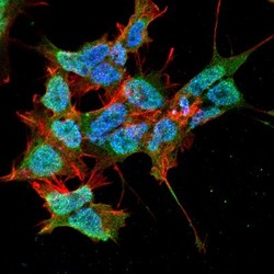
- Immunocytochemistry/Immunofluorescence: PEX6 Antibody (S233-8) [NBP2-42199] - Tissue: Neuroblastoma cell line SK-N-BE. Species: Human. Fixation: 4% Formaldehyde for 15 min at RT. Primary Antibody: Mouse Anti-PEX6 Monoclonal Antibody at 1:100 for 60 min at RT. Secondary Antibody: Goat Anti-Mouse ATTO 488 at 1:100 for 60 min at RT. Counterstain: Phalloidin Texas Red F-Actin stain; DAPI (blue) nuclear stain at 1:1000; 1:5000 for 60 min RT, 5 min RT. Localization: Cytoplasm, Peroxisome, Peroxisome Membrane, Nucleus. Magnification: 60X.
