Antibody data
- Antibody Data
- Antigen structure
- References [3]
- Comments [0]
- Validations
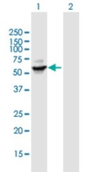
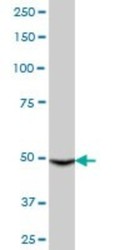
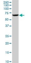
- Western blot [3]
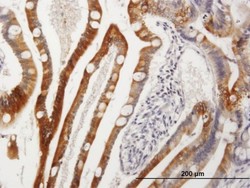
- Immunohistochemistry [1]
Submit
Validation data
Reference
Comment
Report error
- Product number
- H00001576-B01P - Provider product page

- Provider
- Novus Biologicals
- Proper citation
- Novus Cat#H00001576-B01P, RRID:AB_2090340
- Product name
- Mouse Polyclonal Cytochrome P450 3A4 Antibody
- Antibody type
- Polyclonal
- Description
- Protein A purified. CYP3A4 - cytochrome P450, family 3, subfamily A, polypeptide 4,
- Reactivity
- Human, Mouse, Canine
- Host
- Mouse
- Isotype
- IgG
- Vial size
- 0.05 mg
- Storage
- Aliquot and store at -20C or -80C. Avoid freeze-thaw cycles.
Submitted references Dermal xenobiotic metabolism: a comparison between native human skin, four in vitro skin test systems and a liver system.
Phase I and II metabolism and MRP2-mediated export of bosentan in a MDCKII-OATP1B1-CYP3A4-UGT1A1-MRP2 quadruple-transfected cell line.
Oncostatin m renders epithelial cell adhesion molecule-positive liver cancer stem cells sensitive to 5-Fluorouracil by inducing hepatocytic differentiation.
Wiegand C, Hewitt NJ, Merk HF, Reisinger K
Skin pharmacology and physiology 2014;27(5):263-75
Skin pharmacology and physiology 2014;27(5):263-75
Phase I and II metabolism and MRP2-mediated export of bosentan in a MDCKII-OATP1B1-CYP3A4-UGT1A1-MRP2 quadruple-transfected cell line.
Fahrmayr C, König J, Auge D, Mieth M, Münch K, Segrestaa J, Pfeifer T, Treiber A, Fromm M
British journal of pharmacology 2013 May;169(1):21-33
British journal of pharmacology 2013 May;169(1):21-33
Oncostatin m renders epithelial cell adhesion molecule-positive liver cancer stem cells sensitive to 5-Fluorouracil by inducing hepatocytic differentiation.
Yamashita T, Honda M, Nio K, Nakamoto Y, Yamashita T, Takamura H, Tani T, Zen Y, Kaneko S
Cancer research 2010 Jun 1;70(11):4687-97
Cancer research 2010 Jun 1;70(11):4687-97
No comments: Submit comment
Supportive validation
- Submitted by
- Novus Biologicals (provider)
- Main image

- Experimental details
- Western Blot: Cytochrome P450 3A4 Antibody [H00001576-B01P] - Analysis of CYP3A4 expression in transfected 293T cell line by CYP3A4 polyclonal antibody. Lane 1: CYP3A4 transfected lysate(55.33 KDa). Lane 2: Non-transfected lysate.
- Submitted by
- Novus Biologicals (provider)
- Main image

- Experimental details
- Western Blot: Cytochrome P450 3A4 Antibody [H00001576-B01P] - Analysis of CYP3A4 expression in human liver.
- Submitted by
- Novus Biologicals (provider)
- Main image

- Experimental details
- Western Blot: Cytochrome P450 3A4 Antibody [H00001576-B01P] - Analysis of CYP3A4 expression in HepG2.
Supportive validation
- Submitted by
- Novus Biologicals (provider)
- Main image

- Experimental details
- Immunohistochemistry-Paraffin: Cytochrome P450 3A4 Antibody [H00001576-B01P] - Analysis of purified antibody to CYP3A4 on formalin-fixed paraffin-embedded human small Intestine. (antibody concentration 3 ug/ml)
 Explore
 Validate
 Learn
Learn Western blot