Explore
 Validate
 Learn
LearnPA5-18042
antibody from Invitrogen Antibodies
Targeting: DAP3
bMRP-10, DAP-3, DKFZp686G12159, MGC126058, MGC126059, MRP-S29, MRPS29
 Western blot
Antibody data
- Antibody Data
- Antigen structure
- References [0]
- Comments [0]
- Validations
- Western blot [3]
- Immunocytochemistry [1]
- Immunohistochemistry [2]
Submit
Validation data
Reference
Comment
Report error
- Product number
- PA5-18042 - Provider product page

- Provider
- Invitrogen Antibodies
- Product name
- DAP3 Polyclonal Antibody
- Antibody type
- Polyclonal
- Antigen
- Synthetic peptide
- Description
- This antibody is tested in Peptide ELISA: antibody detection limit dilution 64,000.
- Reactivity
- Human
- Host
- Goat
- Isotype
- IgG
- Vial size
- 100 µg
- Concentration
- 0.5 mg/mL
- Storage
- -20° C, Avoid Freeze/Thaw Cycles
No comments: Submit comment
Supportive validation
- Submitted by
- Invitrogen Antibodies (provider)
- Main image

- Experimental details
- Western blot of HEK293 overexpressing DAP3 using Product # PA5-18042, mock transfection as a control in first lane.

- Submitted by
- Invitrogen Antibodies (provider)
- Main image

- Experimental details
- Western Blot staining of HeLa cell lysate using Product # PA5-18042 at a concentration of 3 µg/mL, the primary antibody incubation was 1 hour and the detection method was chemiluminescence.

- Submitted by
- Invitrogen Antibodies (provider)
- Main image

- Experimental details
- Western blot of HEK293 overexpressing DAP3 using Product # PA5-18042, mock transfection as a control in first lane.

Supportive validation
- Submitted by
- Invitrogen Antibodies (provider)
- Main image

- Experimental details
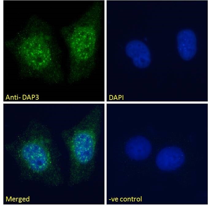
- Immunocytochemical analysis of DAP3 in MCF7 cells using a DAP3 polyclonal antibody (Product #PA5-18042). MCF7 cells were permeabilized with 0.15% Triton. Cells were incubated with 10 µg/mL of primary antibody for one hour followed by an Alexa Fluor 488 secondary antibody at a concentration of 2 µg/mL. Nuclear staining can be seen as shown above. The nuclear stain is DAPI (blue). Negative control: Unimmunized goat IgG (10 µg/mL) followed by an Alexa Fluor 488 secondary antibody (2 µg/mL).
Supportive validation
- Submitted by
- Invitrogen Antibodies (provider)
- Main image

- Experimental details
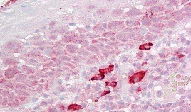
- Immunohistochemical analysis of DAP3 in Human Cortex using a DAP3 monoclonal antibody (Product #PA5-18042) at 2.5 µg/mL. The Human Cortex tissue section was paraffin embeded and detected using steamed antigen retrieval with citrate buffer pH 6, AP-staining.

- Submitted by
- Invitrogen Antibodies (provider)
- Main image

- Experimental details
- Immunohistochemical analysis of DAP3 in Human Tonsil using a DAP3 monoclonal antibody (Product #PA5-18042) at 2.5 µg/mL. The Human Tonsil tissue section was paraffin embeded and detected using steamed antigen retrieval with citrate buffer pH 6, AP-staining.