Explore
 Validate
 Learn
Learn Western blot
Antibody data
- Antibody Data
- Antigen structure
- References [1]
- Comments [0]
- Validations
- Western blot [4]
- Immunocytochemistry [1]
Submit
Validation data
Reference
Comment
Report error
- Product number
- 703154 - Provider product page

- Provider
- Invitrogen Antibodies
- Product name
- TBK1 Recombinant Rabbit Monoclonal Antibody (12H60L39)
- Antibody type
- Monoclonal
- Antigen
- Synthetic peptide
- Reactivity
- Human, Mouse
- Host
- Rabbit
- Isotype
- IgG
- Antibody clone number
- 12H60L39
- Vial size
- 100 µg
- Concentration
- 0.5 mg/mL
- Storage
- Store at 4°C short term. For long term storage, store at -20°C, avoiding freeze/thaw cycles.
Submitted references The A137R Protein of African Swine Fever Virus Inhibits Type I Interferon Production via the Autophagy-Mediated Lysosomal Degradation of TBK1.
Sun M, Yu S, Ge H, Wang T, Li Y, Zhou P, Pan L, Han Y, Yang Y, Sun Y, Li S, Li LF, Qiu HJ
Journal of virology 2022 May 11;96(9):e0195721
Journal of virology 2022 May 11;96(9):e0195721
No comments: Submit comment
Supportive validation
- Submitted by
- Invitrogen Antibodies (provider)
- Main image

- Experimental details
- Knockdown of TBK1 was achieved by transfecting K-562 cells with TBK1 specific validated siRNA (Silencer® select Product # s761). Western blot analysis (Fig a) was performed using whole cell extracts from TBK1 knockdown cells (Lane 3), non-specific scrambled siRNA transfected cells (Lane 2) and untransfected cells (Lane 1). The blot was probed with Anti-TBK1 Recombinant Rabbit Monoclonal Antibody (Product # 703154, 1:1000 dilution) and Goat anti-Rabbit IgG (H+L) Superclonal™ Secondary Antibody, HRP conjugate (Product # A27036, 0.25 µg/mL, 1:10000 dilution). Densitometric analysis of this western blot is shown in the histogram (Fig b). Loss of signal upon siRNA mediated knockdown confirms that antibody is specific to TBK1.

- Submitted by
- Invitrogen Antibodies (provider)
- Main image
- Experimental details
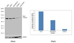
- Western blot was performed using Anti-TBK1 Recombinant Rabbit Monoclonal Antibody (Product # 703154) and a ~84 kDa band corresponding to TBK1 was observed across the cell lines tested. Whole cell extracts (40 µg lysate) of K562 (Lane 1), MCF7 (Lane 2), MDA-MB-231 (Lane 3), SK-BR-3 (Lane 4) and tissue extracts of Mouse Brain (Lane 5) were electrophoresed using Novex® NuPAGE® 4-12% Bis-Tris gel (Product # NP0322BOX). Resolved proteins were then transferred onto a nitrocellulose membrane by iBlot® 2 Dry Blotting System (Product # IB21001). The blot was probed with the primary antibody (1:1000 dilution) and detected by chemiluminescence using Goat anti-Rabbit IgG (H+L) Superclonal™ Secondary Antibody, HRP conjugate (Product # A27036, 1:10000 dilution) using the iBright FL 1500 (Product # A44115). Chemiluminescent detection was performed using Novex® ECL Chemiluminescent Substrate Reagent Kit (Product # WP20005).

- Submitted by
- Invitrogen Antibodies (provider)
- Main image

- Experimental details
- CRISPR-Cas9 mediated genome editing ofTBK1 (as confirmed by next generation sequencing) was achieved by using LentiArray™ Lentiviral sgRNA (Product # A32042, AssayID CRISPR1012237_LV) and LentiArray Cas9 Lentivirus (Product # A32064). Fig (a) Western blot analysis of TBK1 was performed by loading 30 µg of A549 wild type (Lane 1), A549 Cas9 (Lane 2) and A549 Cas9 cells transduced with TBK1 Lentiviral sgRNA (Lane 3) whole cell extracts. The samples were electrophoresed using NuPAGE™ Novex™ 4-12% Bis-Tris Protein Gel (Product # NP0321BOX). Resolved proteins were then transferred onto a nitrocellulose membrane (Product # IB23001) by iBlot® 2 Dry Blotting System (Product # IB21001). The blot was probed with Anti-TBK1 Recombinant Rabbit Monoclonal Antibody (12H60L39) (Product # 703154) using 1:1000 dilution and Goat anti-Rabbit IgG (H+L) Superclonal™ Recombinant Secondary Antibody, HRP (Product # A27036 1:6000 dilution).Chemiluminescent detection was performed using Novex® ECL Chemiluminescent Substrate Reagent Kit (Product # WP20005). A loss of signal in sgRNA transduced cells using the LentiArray™ CRISPR product line confirms that antibody is specific toTBK1 (Fig (b)). Uncharacterized bands were observed at ~12 kDa and ~20 kDa in all the samples.

- Submitted by
- Invitrogen Antibodies (provider)
- Main image

- Experimental details
- Western blot analysis of TBK1 was performed by loading 10 µg of WT (lane 1) and TBK1 CRISPR KO (lane 2) U2OS cell lysates in RIPA buffer onto a 4-15% gradient polyacrylamide gel. Proteins were transferred to nitrocellulose membrane and blocked in 5% milk. Ponceau stained transfer of blot is shown. TBK1 was detected using a TBK1 recombinant monoclonal antibody (Product # 703154) at a dilution of 1:5,000 in 5% BSA in TBST overnight at 4 deg, followed by secondary antibody diluted to 0.2 µg/mL using Goat anti-Rabbit IgG (H+L) HRP antibody (Product # 65-6120). Chemiluminescent detection was performed using Pierce ECL Western Blotting Substrate (Product # 32106). Data courtesy of YCharOS Inc., an open science company with the mission of characterizing commercially available antibodies using knockout validation.

Supportive validation
- Submitted by
- Invitrogen Antibodies (provider)
- Main image

- Experimental details
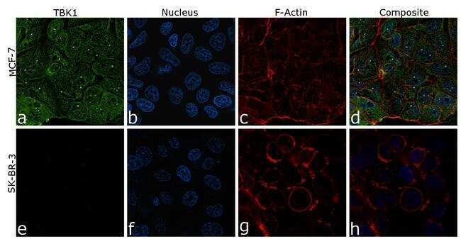
- For immunofluorescence analysis, MCF-7 and SK-BR-3 cells were fixed and permeabilized for detection of endogenous TBK1 using Anti-TBK1 Recombinant Rabbit Monoclonal Antibody (Product # 703154, 1:100) and labeled with Goat anti-Rabbit IgG (H+L) Highly Cross-Adsorbed Secondary Antibody, Alexa Fluor Plus 488 conjugate (Product # A32731, 1:2000). Nuclei (blue) were stained using ProLong™ Diamond Antifade Mountant with DAPI (Product # P36962), and Rhodamine Phalloidin (Product # R415, 1:800) was used for cytoskeletal F-actin (red) staining. Panel a-d) clearly demonstrates the nuclear and cytoplasmic localization of TBK1 in MDA-MB-231 cells. Panel e-h) shows reduced specific staining in SK-BR-3 cells which has low expression compared to MDA-MB-231, demonstrating specificity. The images were captured at 60X magnification.