 Explore
 Validate
 Learn
Learn Immunocytochemistry
 Immunohistochemistry
Antibody data
- Antibody Data
- Antigen structure
- References [0]
- Comments [0]
- Validations
- Immunocytochemistry [1]
- Flow cytometry [2]
Submit
Validation data
Reference
Comment
Report error
- Product number
- MA1-10175 - Provider product page

- Provider
- Invitrogen Antibodies
- Product name
- CD3 Monoclonal Antibody (OKT3)
- Antibody type
- Monoclonal
- Antigen
- Other
- Description
- This antibody recognizes an extracellular epitope on CD3 antigen of the TCR/CD3 complex on mature human T cells. This antibody, also known as Orthoclone OKT3 or Muromonab-CD3, has been extensively used as a drug for therapy of acute, glucocorticoid resistant rejection of allogenic renal, heart and liver transplants. It has also been investigated for use in treating T-cell acute lymphoblastic leukemia.
- Reactivity
- Human
- Host
- Mouse
- Isotype
- IgG
- Antibody clone number
- OKT3
- Vial size
- 100 µg
- Concentration
- 1 mg/mL
- Storage
- 4° C, do not freeze
No comments: Submit comment
Supportive validation
- Submitted by
- Invitrogen Antibodies (provider)
- Main image

- Experimental details
- Immunofluorescence analysis of CD3e was performed using 70% confluent log phase Jurkat cells. The cells were fixed with 4% paraformaldehyde for 10 minutes, permeabilized with 0.1% Triton X-100 for 10 minutes, and blocked with 1% BSA for 1 hour at room temperature. The cells were labeled with CD3e (OKT3) Mouse Monoclonal Antibody (Product # MA1-10175) at 2 µg/mL in 0.1% BSA and incubated for 3 hours at room temperature and then labeled with Goat anti-Mouse IgG (H+L) Superclonal Secondary Antibody, Alexa Fluor® 488 conjugate (Product # A28175) at a dilution of 1:2000 for 45 minutes at room temperature (Panel a: green). Nuclei (Panel b: blue) were stained with SlowFade® Gold Antifade Mountant with DAPI (Product # S36938). F-actin (Panel c: red) was stained with Alexa Fluor® 555 Rhodamine Phalloidin (Product # R415, 1:300). Panel d represents the merged image showing membranous and cytoplasmic localization. Panel e shows the no primary antibody control. The images were captured at 60X magnification.

Supportive validation
- Submitted by
- Invitrogen Antibodies (provider)
- Main image

- Experimental details
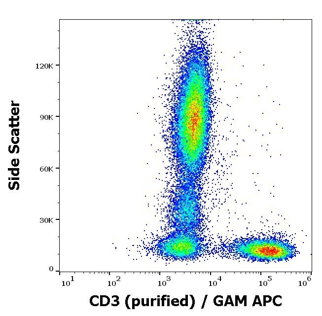
- Flow cytometry surface staining pattern of human peripheral whole blood stained using anti-human CD3 (OKT3) purified Monoclonal antibody (Product # MA1-10175) (concentration in sample 1 µg/mL) GAM APC.
- Submitted by
- Invitrogen Antibodies (provider)
- Main image

- Experimental details
- Separation of human CD3 positive lymphocytes (red-filled) from neutrophil granulocytes (black-dashed) in flow cytometry analysis (surface staining) of human peripheral whole blood stained using anti-human CD3 (OKT3) purified Monoclonal antibody (Product # MA1-10175) (concentration in sample 1 µg/mL) GAM APC.
