Explore
 Validate
 Learn
Learn Western blot
Antibody data
- Antibody Data
- Antigen structure
- References [6]
- Comments [0]
- Validations
- Western blot [1]
- Immunohistochemistry [1]
- Flow cytometry [1]
Submit
Validation data
Reference
Comment
Report error
- Product number
- SM7100P - Provider product page

- Provider
- Acris Antibodies GmbH
- Proper citation
- Acris Antibodies GmbH Cat#SM7100P, RRID:AB_1008040
- Product name
- anti CD282 / TLR2
- Antibody type
- Monoclonal
- Antigen
- Extracellular domain of human TLR2
- Reactivity
- Human
- Host
- Mouse
- Isotype
- IgG
- Antibody clone number
- 1030A5.138
- Vial size
- 0.2 ml
Submitted references Differential expression of TLR-2 and TLR-4 in the epithelial cells in oral lichen planus.
TLR2 expression is regulated by microRNA miR-19 in rheumatoid fibroblast-like synoviocytes.
Inhibition of neutrophil apoptosis by TLR agonists in whole blood: involvement of the phosphoinositide 3-kinase/Akt and NF-kappaB signaling pathways, leading to increased levels of Mcl-1, A1, and phosphorylated Bad.
Microarray analysis reveals induction of lipoprotein genes in mucoid Pseudomonas aeruginosa: implications for inflammation in cystic fibrosis.
Soluble forms of Toll-like receptor (TLR)2 capable of modulating TLR2 signaling are present in human plasma and breast milk.
Glucocorticoids synergistically enhance nontypeable Haemophilus influenzae-induced Toll-like receptor 2 expression via a negative cross-talk with p38 MAP kinase.
Janardhanam SB, Prakasam S, Swaminathan VT, Kodumudi KN, Zunt SL, Srinivasan M
Archives of oral biology 2012 May;57(5):495-502
Archives of oral biology 2012 May;57(5):495-502
TLR2 expression is regulated by microRNA miR-19 in rheumatoid fibroblast-like synoviocytes.
Philippe L, Alsaleh G, Suffert G, Meyer A, Georgel P, Sibilia J, Wachsmann D, Pfeffer S
Journal of immunology (Baltimore, Md. : 1950) 2012 Jan 1;188(1):454-61
Journal of immunology (Baltimore, Md. : 1950) 2012 Jan 1;188(1):454-61
Inhibition of neutrophil apoptosis by TLR agonists in whole blood: involvement of the phosphoinositide 3-kinase/Akt and NF-kappaB signaling pathways, leading to increased levels of Mcl-1, A1, and phosphorylated Bad.
François S, El Benna J, Dang PM, Pedruzzi E, Gougerot-Pocidalo MA, Elbim C
Journal of immunology (Baltimore, Md. : 1950) 2005 Mar 15;174(6):3633-42
Journal of immunology (Baltimore, Md. : 1950) 2005 Mar 15;174(6):3633-42
Microarray analysis reveals induction of lipoprotein genes in mucoid Pseudomonas aeruginosa: implications for inflammation in cystic fibrosis.
Firoved AM, Ornatowski W, Deretic V
Infection and immunity 2004 Sep;72(9):5012-8
Infection and immunity 2004 Sep;72(9):5012-8
Soluble forms of Toll-like receptor (TLR)2 capable of modulating TLR2 signaling are present in human plasma and breast milk.
LeBouder E, Rey-Nores JE, Rushmere NK, Grigorov M, Lawn SD, Affolter M, Griffin GE, Ferrara P, Schiffrin EJ, Morgan BP, Labéta MO
Journal of immunology (Baltimore, Md. : 1950) 2003 Dec 15;171(12):6680-9
Journal of immunology (Baltimore, Md. : 1950) 2003 Dec 15;171(12):6680-9
Glucocorticoids synergistically enhance nontypeable Haemophilus influenzae-induced Toll-like receptor 2 expression via a negative cross-talk with p38 MAP kinase.
Shuto T, Imasato A, Jono H, Sakai A, Xu H, Watanabe T, Rixter DD, Kai H, Andalibi A, Linthicum F, Guan YL, Han J, Cato AC, Lim DJ, Akira S, Li JD
The Journal of biological chemistry 2002 May 10;277(19):17263-70
The Journal of biological chemistry 2002 May 10;277(19):17263-70
No comments: Submit comment
Supportive validation
- Submitted by
- Acris Antibodies GmbH (provider)
- Main image

- Experimental details
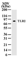
- Western blot analysis of TLR2 in 30 ugs of Ramos whole cell lysate using SM7100P at 1/500.
Supportive validation
- Submitted by
- Acris Antibodies GmbH (provider)
- Main image
- Experimental details
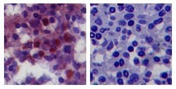
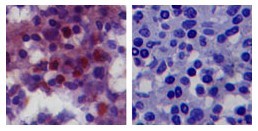
- IHC analysis of TLR2 in paraffin-embedded human spleen tissue using SM7100P (left) and isotype control (right).
Supportive validation
- Submitted by
- Acris Antibodies GmbH (provider)
- Main image

- Experimental details
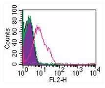
- Cell surface flow analysis of TLR2 in 10e6 human monocytes using 2.5 µl of SM7100P. The shaded histogram represents cells with no antibody present, Green represents Mouse IgG1 Isotype Control Cat.-No AM33027AF-N, Red represents TLR2 Antibody. Goat anti Mouse IgG1 PE secondary was used.