Explore
 Validate
 Learn
Learn Western blot
 Immunocytochemistry
Antibody data
- Antibody Data
- Antigen structure
- References [2]
- Comments [0]
- Validations
- Immunocytochemistry [3]
- Immunohistochemistry [3]
Submit
Validation data
Reference
Comment
Report error
- Product number
- GTX22803 - Provider product page

- Provider
- GeneTex
- Proper citation
- GeneTex Cat#GTX22803, RRID:AB_384860
- Product name
- AChE antibody [HR2]
- Antibody type
- Monoclonal
- Reactivity
- Human, Mouse, Bovine, Feline, Guinea Pig, Rabbit
- Host
- Mouse
Submitted references In vivo localization of human acetylcholinesterase-derived species in a β-sheet conformation at the core of senile plaques in Alzheimer's disease.
Immunopurification of Acetylcholinesterase from Red Blood Cells for Detection of Nerve Agent Exposure.
Jean L, Brimijoin S, Vaux DJ
The Journal of biological chemistry 2019 Apr 19;294(16):6253-6272
The Journal of biological chemistry 2019 Apr 19;294(16):6253-6272
Immunopurification of Acetylcholinesterase from Red Blood Cells for Detection of Nerve Agent Exposure.
Dafferner AJ, Schopfer LM, Xiao G, Cashman JR, Yerramalla U, Johnson RC, Blake TA, Lockridge O
Chemical research in toxicology 2017 Oct 16;30(10):1897-1910
Chemical research in toxicology 2017 Oct 16;30(10):1897-1910
No comments: Submit comment
Supportive validation
- Submitted by
- GeneTex (provider)
- Main image
- Experimental details
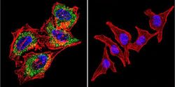
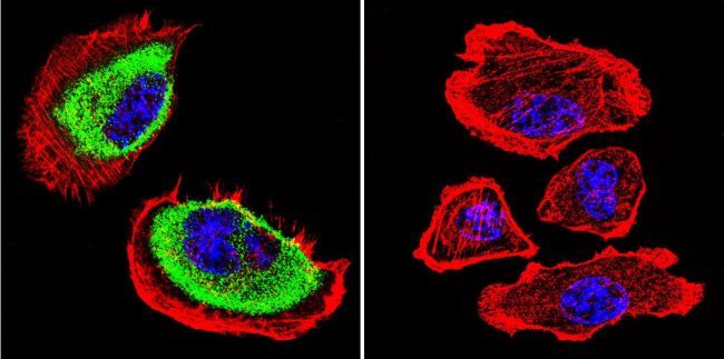
- Immunofluorescent analysis of Acetylcholinesterase in HeLa cells. Acetylcholinesterase staining (green), F-Actin staining with Phalloidin (red) and nuclei with DAPI (blue) is shown. Cells were grown on slides and fixed with formaldehyde prior to staining. Cells were probed without (control) or with Acetylcholinesterase antibody [HR2] at a dilution of 1:200 over night at 4¢XC, washed with PBS and incubated with a proper secondary antibody. Images were taken at 60X magnification.
- Submitted by
- GeneTex (provider)
- Main image
- Experimental details
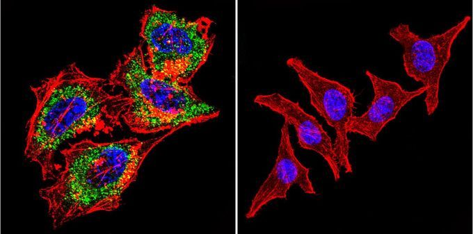
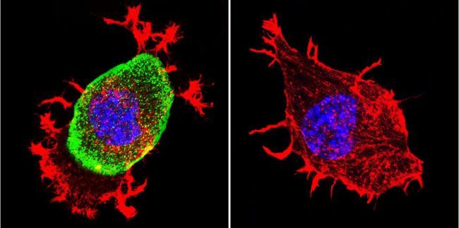
- Immunofluorescent analysis of Acetylcholinesterase in U251 Cells. Acetylcholinesterase staining (green), F-Actin staining with Phalloidin (red) and nuclei with DAPI (blue) is shown. Cells were grown on chamber slides and fixed with formaldehyde prior to staining. Cells were probed without (control) or with Acetylcholinesterase antibody [HR2] at a dilution of 1:200 over night at 4¢XC, washed with PBS and incubated with a proper secondary antibody. Images were taken at 60X magnification.
- Submitted by
- GeneTex (provider)
- Main image

- Experimental details
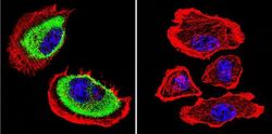
- Immunofluorescent analysis of Acetylcholinesterase in Neuro-2a cells. Acetylcholinesterase staining (green), F-Actin staining with Phalloidin (red) and nuclei with DAPI (blue) is shown. Cells were grown on chamber slides and fixed with formaldehyde prior to staining. Cells were probed without (control) or with Acetylcholinesterase antibody [HR2] at a dilution of 1:200 over night at 4¢XC, washed with PBS and incubated with a proper secondary antibody. Images were taken at 60X magnification.
Supportive validation
- Submitted by
- GeneTex (provider)
- Main image
- Experimental details
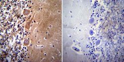
- Immunohistochemistry was performed on normal deparaffinized human Cerebellum tissue. To expose target proteins, heat induced antigen retrieval was performed using 10mM sodium citrate (pH6.0) buffer, microwaved for 8-15 minutes. Following antigen retrieval tissues were blocked in 3% BSA-PBS for 30 minutes at room temperature. Tissues were then probed with or without Acetylcholinesterase antibody [HR2] at a dilution of 1:50 overnight at 4¢XC in a humidified chamber. Tissues were washed extensively with PBST and endogenous peroxidase activity was quenched with a peroxidase suppressor. Detection was performed using a biotin-conjugated secondary antibody and SA-HRP, followed by colorimetric detection using DAB. Tissues were counterstained with hematoxylin and prepped for mounting.

- Submitted by
- GeneTex (provider)
- Main image
- Experimental details
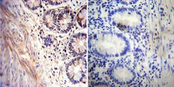
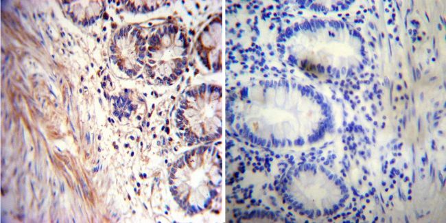
- Immunohistochemistry was performed on normal deparaffinized human Rectum tissue. To expose target proteins, heat induced antigen retrieval was performed using 10mM sodium citrate (pH6.0) buffer, microwaved for 8-15 minutes. Following antigen retrieval tissues were blocked in 3% BSA-PBS for 30 minutes at room temperature. Tissues were then probed with or without Acetylcholinesterase antibody [HR2] at a dilution of 1:20 overnight at 4¢XC in a humidified chamber. Tissues were washed extensively with PBST and endogenous peroxidase activity was quenched with a peroxidase suppressor. Detection was performed using a biotin-conjugated secondary antibody and SA-HRP, followed by colorimetric detection using DAB. Tissues were counterstained with hematoxylin and prepped for mounting.
- Submitted by
- GeneTex (provider)
- Main image

- Experimental details
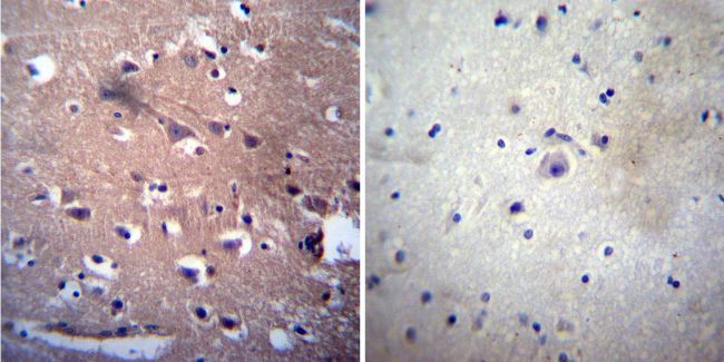
- Immunohistochemistry was performed on normal deparaffinized human Brain tissue. To expose target proteins, heat induced antigen retrieval was performed using 10mM sodium citrate (pH6.0) buffer, microwaved for 8-15 minutes. Following antigen retrieval tissues were blocked in 3% BSA-PBS for 30 minutes at room temperature. Tissues were then probed with or without Acetylcholinesterase antibody [HR2] at a dilution of at a dilution of 1:200 overnight at 4¢XC in a humidified chamber. Tissues were washed extensively with PBST and endogenous peroxidase activity was quenched with a peroxidase suppressor. Detection was performed using a biotin-conjugated secondary antibody and SA-HRP, followed by colorimetric detection using DAB. Tissues were counterstained with hematoxylin and prepped for mounting.