Explore
 Validate
 Learn
Learn Western blot
 ELISA
ELISAAntibody data
- Antibody Data
- Antigen structure
- References [1]
- Comments [0]
- Validations
- Western blot [3]
- Flow cytometry [2]
Submit
Validation data
Reference
Comment
Report error
- Product number
- NB100-1519 - Provider product page

- Provider
- Novus Biologicals
- Proper citation
- Novus Cat#NB100-1519, RRID:AB_2242347
- Product name
- Goat Polyclonal Acetylcholinesterase/ACHE Antibody
- Antibody type
- Polyclonal
- Description
- Immunogen affinity purified. This antibody is expected to recognise isoform NP_000656 only (the ubiquitously expressed, hydrophillic form).
- Reactivity
- Human, Mouse, Rat
- Host
- Goat
- Isotype
- IgG
- Vial size
- 0.1 mg
- Concentration
- 0.5 mg/ml
- Storage
- Store at -20C. Avoid freeze-thaw cycles.
Submitted references The intact human acetylcholinesterase C-terminal oligomerization domain is alpha-helical in situ and in isolation, but a shorter fragment forms beta-sheet-rich amyloid fibrils and protofibrillar oligomers.
Cottingham MG, Voskuil JL, Vaux DJ
Biochemistry 2003 Sep 16;42(36):10863-73
Biochemistry 2003 Sep 16;42(36):10863-73
No comments: Submit comment
Supportive validation
- Submitted by
- Novus Biologicals (provider)
- Main image
- Experimental details
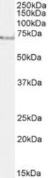
- Western Blot: Acetylcholinesterase/ACHE Antibody [NB100-1519] - (0.3ug/ml) staining of Rat Brain lysate (35ug protein in RIPA buffer). Detected by chemiluminescence.
- Submitted by
- Novus Biologicals (provider)
- Main image

- Experimental details
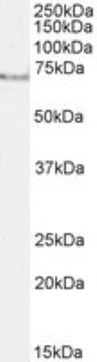
- Western Blot: Acetylcholinesterase/ACHE Antibody [NB100-1519] - (0.3ug/ml) staining of Daudi (A), HeLa (B) and Jurkat (C) cell lysate (35ug protein in RIPA buffer). Detected by chemiluminescence.
- Submitted by
- Novus Biologicals (provider)
- Main image
- Experimental details
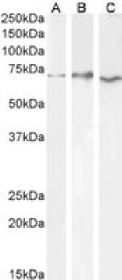
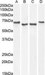
- Western Blot: Acetylcholinesterase/ACHE Antibody [NB100-1519] - Staining of HepG2 (A) and Daudi cell lysates with antibody at 1 ug/mL (B) and HeLa (C) and Jurkat (D) cell lysates with antibody at 0.3 ug/mL (35 ug protein in RIPA buffer). Detected by chemiluminescence.

Supportive validation
- Submitted by
- Novus Biologicals (provider)
- Main image
- Experimental details
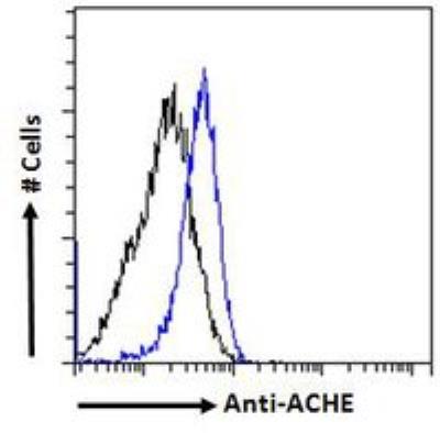
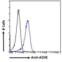
- Flow Cytometry: Acetylcholinesterase/ACHE Antibody [NB100-1519] - analysis of paraformaldehyde fixed HeLa cells (blue line), permeabilized with 0.5% Triton. Primary incubation 1hr (10ug/ml) followed by Alexa Fluor 488 secondary antibody (2ug/ml). IgG control: Unimmunized goat IgG (black line) followed by Alexa Fluor 488 secondary antibody.
- Submitted by
- Novus Biologicals (provider)
- Main image
- Experimental details
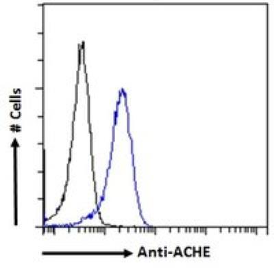
- Flow Cytometry: Acetylcholinesterase/ACHE Antibody [NB100-1519] - Flow cytometric analysis of paraformaldehyde fixed HeLa cells (blue line), permeabilized with 0.5% Triton. Primary incubation 1hr (10 ug/mL) followed by Alexa Fluor 488 secondary antibody (1 ug/mL). IgG control: Unimmunized goat IgG (black line) followed by Alexa Fluor 488 secondary antibody.