Explore
 Validate
 Learn
LearnHPA006983
antibody from Atlas Antibodies
Targeting: PRDX6
1-Cys, aiPLA2, AOP2, KIAA0106, MGC46173, NSGPx, p29, PRX
 Western blot
Antibody data
- Antibody Data
- Antigen structure
- References [1]
- Comments [0]
- Validations
- Western blot [2]
- Immunohistochemistry [1]
Submit
Validation data
Reference
Comment
Report error
- Product number
- HPA006983 - Provider product page

- Provider
- Atlas Antibodies
- Proper citation
- Atlas Antibodies Cat#HPA006983, RRID:AB_1079595
- Product name
- Anti-PRDX6
- Antibody type
- Polyclonal
- Reactivity
- Human
- Host
- Rabbit
- Conjugate
- Unconjugated
- Antigen sequence
GGLLLGDVAPNFEANTTVGRIRFHDFLGDSWGILF
SHPRDFTPVCTTELGRAAKLAPEFAKRNVKLIALS
IDSVEDHLAWSKDINAYNCEEPTEKLPFPIIDDRN
RELAILLGMLDPAEKDEKGMPVTARVVFVFGPDKK
LKLSILYPA- Isotype
- IgG
- Vial size
- 100 µl
- Storage
- Store at +4°C for short term storage. Long time storage is recommended at -20°C.
Submitted references Human Protein Atlas of redox systems — What can be learnt?
Dammeyer P, Arnér E
Biochimica et Biophysica Acta (BBA) - General Subjects 2011 January;1810(1):111-138
Biochimica et Biophysica Acta (BBA) - General Subjects 2011 January;1810(1):111-138
No comments: Submit comment
Supportive validation
Supportive validation
- Submitted by
- Atlas Antibodies (provider)
- Enhanced method
- Orthogonal validation
- Main image

- Experimental details
- Western blot analysis in human cell lines PC-3 and MCF-7 using Anti-PRDX6 antibody. Corresponding PRDX6 RNA-seq data are presented for the same cell lines. Loading control: Anti-COX4I1.

Supportive validation
- Submitted by
- Atlas Antibodies (provider)
- Main image
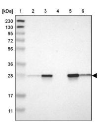
- Experimental details
- Lane 1: Marker [kDa] 230, 130, 95, 72, 56, 36, 28, 17, 11Lane 2: Human cell line RT-4Lane 3: Human cell line U-251MG spLane 4: Human plasma (IgG/HSA depleted)Lane 5: Human liver tissueLane 6: Human tonsil tissue

Supportive validation
- Submitted by
- Atlas Antibodies (provider)
- Main image

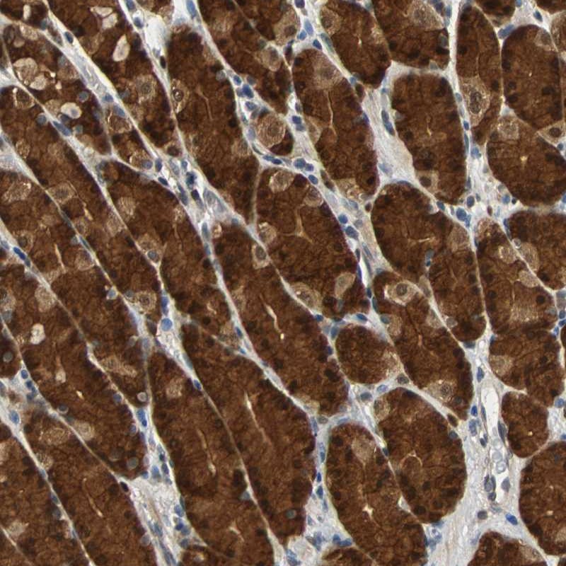
- Experimental details
- Immunohistochemical staining of human stomach shows strong nuclear and cytoplasmic positivity in glandular cells.
- Sample type
- HUMAN