 Explore
 Validate
 Learn
LearnPA5-21882
antibody from Invitrogen Antibodies
Targeting: NUP62
DKFZp547L134, FLJ20822, FLJ43869, IBSN, MGC841, p62, SNDI
 Western blot
Antibody data
- Antibody Data
- Antigen structure
- References [1]
- Comments [0]
- Validations
- Western blot [7]
- Immunocytochemistry [2]
- Immunohistochemistry [2]
- Other assay [1]
Submit
Validation data
Reference
Comment
Report error
- Product number
- PA5-21882 - Provider product page

- Provider
- Invitrogen Antibodies
- Product name
- NUP62 Polyclonal Antibody
- Antibody type
- Polyclonal
- Antigen
- Recombinant protein fragment
- Description
- Recommended positive controls: HepG2, NIH-3T3, JC, BCL-1, C2C12, PC-12.
- Concentration
- 1 mg/mL
Submitted references Physical confinement during cancer cell migration triggers therapeutic resistance and cancer stem cell-like behavior.
Shen Q, Hill T, Cai X, Bui L, Barakat R, Hills E, Almugaiteeb T, Babu A, Mckernan PH, Zalles M, Battiste JD, Kim YT
Cancer letters 2021 May 28;506:142-151
Cancer letters 2021 May 28;506:142-151
No comments: Submit comment
Supportive validation
- Submitted by
- Invitrogen Antibodies (provider)
- Main image
- Experimental details
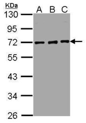
- Western blot analysis of nucleoporin p62 using 30 µg of A) HepG2 (B) MOLT4 and C) Raji lysate. Samples were loaded onto a 10% SDS-PAGE gel and probed with a nucleoporin p62 polyclonal antibody (Product # PA5-21882) at a dilution of 1:5000.

- Submitted by
- Invitrogen Antibodies (provider)
- Main image

- Experimental details
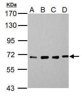
- Western Blot using NUP62 Polyclonal Antibody (Product # PA5-21882). Sample (30 µg of whole cell lysate). Lane A: NIH-3T3. Lane B: JC. Lane C: BCL-1. Lane D: C2C12. 7.5% SDS PAGE. NUP62 Polyclonal Antibody (Product # PA5-21882) diluted at 1:5,000. The HRP-conjugated anti-rabbit IgG antibody was used to detect the primary antibody.

- Submitted by
- Invitrogen Antibodies (provider)
- Main image

- Experimental details
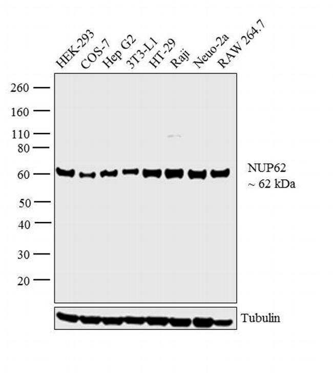
- Western blot analysis was performed on whole cell extracts (30 µg lysate) of HEK-293 (Lane 1), COS-7 (Lane 2), Hep G2 (Lane 3), 3T3-L1 (Lane 4), HT-29 (Lane 5), Raji (Lane 6), Neuro-2a (Lane 7) and RAW 264.7 (Lane 8). The blot was probed with Anti-NUP62 Polyclonal Antibody (Product # PA5-21882, 1:5000 dilution) and detected by chemiluminescence using Goat anti-Rabbit IgG (H+L) Superclonal™ Secondary Antibody, HRP conjugate (Product # A27036, 0.25 ug/ml, 1:4000 dilution). A 62 kDa band corresponding to NUP62 was observed across the cell lines tested.
- Submitted by
- Invitrogen Antibodies (provider)
- Main image

- Experimental details
- Western Blot analysis of NUP62 was performed by separating 30 µg of whole cell extracts by 10% SDS-PAGE. Proteins were transferred to a membrane and probed with a NUP62 Polyclonal Antibody (Product # PA5-21882) at a dilution of 1:5000. The HRP-conjugated anti-rabbit IgG antibody was used to detect the primary antibody.

- Submitted by
- Invitrogen Antibodies (provider)
- Main image

- Experimental details
- Western Blot using NUP62 Polyclonal Antibody (Product # PA5-21882). Sample (30 µg of whole cell lysate). Lane A: PC-12. 7.5% SDS PAGE. NUP62 Polyclonal Antibody (Product # PA5-21882) diluted at 1:5,000. The HRP-conjugated anti-rabbit IgG antibody was used to detect the primary antibody.

- Submitted by
- Invitrogen Antibodies (provider)
- Main image

- Experimental details
- Western Blot using NUP62 Polyclonal Antibody (Product # PA5-21882). Sample (30 µg of whole cell lysate). Lane A: NIH-3T3. Lane B: JC. Lane C: BCL-1. Lane D: C2C12. 7.5% SDS PAGE. NUP62 Polyclonal Antibody (Product # PA5-21882) diluted at 1:5,000. The HRP-conjugated anti-rabbit IgG antibody was used to detect the primary antibody.
- Submitted by
- Invitrogen Antibodies (provider)
- Main image

- Experimental details
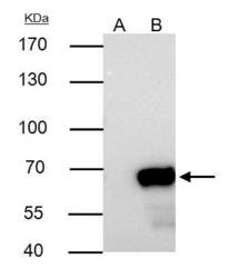
- Knockdown of NUP62 was achieved by transfecting HEK293 cells with NUP62 specific siRNAs (Silencer® select Product # s24247, s24248). Western blot analysis (Fig. a) was performed using whole cell extracts from the NUP62 knockdown cells (lane 3), non-specific scrambled siRNA transfected cells (lane 2) and untransfected cells (lane 1). The blots were probed with NUP62 Polyclonal Antibody (Product # PA5-21882, 1:1000 dilution) and Goat anti-Rabbit IgG (H+L) Superclonal™ Secondary Antibody, HRP conjugate (Product # A27036, 0.25ug/ml, 1:4000 dilution). Densitometric analysis of this western blot is shown in histogram (Fig. b). Loss of signal upon siRNA mediated knock down confirms that antibody is specific to NUP62.

Supportive validation
- Submitted by
- Invitrogen Antibodies (provider)
- Main image

- Experimental details
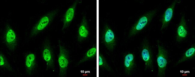
- Immunocytochemistry-Immunofluorescence analysis of NUP62 was performed in HeLa cells fixed in 4% paraformaldehyde at RT for 15 min. Green: NUP62 Polyclonal Antibody (Product # PA5-21882) diluted at 1:750. Blue: Hoechst 33342 staining. Scale bar = 10 µm.
- Submitted by
- Invitrogen Antibodies (provider)
- Main image
- Experimental details
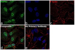
- Immunofluorescence analysis of NUP62 was performed using 70% confluent log phase HepG2 cells. The cells were fixed with 4% paraformaldehyde for 10 minutes, permeabilized with 0.1% Triton™ X-100 for 15 minutes, and blocked with 1% BSA for 1 hour at room temperature. The cells were labeled with NUP62 Rabbit Polyclonal Antibody(Product # PA5-21882) at 5 µg/mL in 0.1% BSA, incubated at 4 degree Celsius overnight and then labeled with Goat anti-Rabbit IgG (H+L) Superclonal™ Secondary Antibody, Alexa Fluor® 488 conjugate (Product # A27034) at a dilution of 1:2000 for 45 minutes at room temperature (Panel a: green). Nuclei (Panel b: blue) were stained with ProLong™ Diamond Antifade Mountant with DAPI (Product # P36962). F-actin (Panel c: red) was stained with Rhodamine Phalloidin (Product # R415, 1:300). Panel d represents the merged image showing Nucleus and cytoplasmic localization. Panel e represents control cells with no primary antibody to assess background. The images were captured at 60X magnification.

Supportive validation
- Submitted by
- Invitrogen Antibodies (provider)
- Main image

- Experimental details
- Immunohistochemical analysis of paraffin-embedded NCIN87 xenograft, using nucleoporin p62 (Product # PA5-21882) antibody at 1:500 dilution. Antigen Retrieval: EDTA based buffer, pH 8.0, 15 min.

- Submitted by
- Invitrogen Antibodies (provider)
- Main image

- Experimental details
- NUP62 Polyclonal Antibody detects Nucleoporin p62 protein at nucleus by immunohistochemical analysis. Sample: Paraffin-embedded rat brain. Nucleoporin p62 stained by NUP62 Polyclonal Antibody (Product # PA5-21882) diluted at 1:500. Antigen Retrieval: Citrate buffer, pH 6.0, 15 min.

Supportive validation
- Submitted by
- Invitrogen Antibodies (provider)
- Main image
- Experimental details
- NUP62 Polyclonal Antibody immunoprecipitates NUP62 protein in IP experiments. IP samples: 293T whole cell extract. A. Control with 4 µg of preimmune Rabbit IgG. B. Immunoprecipitation of NUP62 protein by 4 µg NUP62 Polyclonal Antibody (Product # PA5-21882). 7.5 % SDS-PAGE. The immunoprecipitated NUP62 protein was detected by NUP62 Polyclonal Antibody (Product # PA5-21882) diluted at 1:1,000.
