 Explore
 Validate
 Learn
Learn Western blot
Antibody data
- Antibody Data
- Antigen structure
- References [1]
- Comments [0]
- Validations
- Western blot [1]
- Immunocytochemistry [2]
- Immunohistochemistry [2]
Submit
Validation data
Reference
Comment
Report error
- Product number
- HPA006777 - Provider product page

- Provider
- Atlas Antibodies
- Proper citation
- Atlas Antibodies Cat#HPA006777, RRID:AB_1080507
- Product name
- Anti-USP2
- Antibody type
- Polyclonal
- Description
- Polyclonal Antibody against Human USP2, Gene description: ubiquitin specific peptidase 2, Alternative Gene Names: UBP41, Validated applications: ICC, IHC, WB, Uniprot ID: O75604, Storage: Store at +4°C for short term storage. Long time storage is recommended at -20°C.
- Reactivity
- Human
- Host
- Rabbit
- Conjugate
- Unconjugated
- Isotype
- IgG
- Vial size
- 100 µl
- Concentration
- 0.3 mg/ml
- Storage
- Store at +4°C for short term storage. Long time storage is recommended at -20°C.
Submitted references USP2 Inhibits Lung Cancer Pathogenesis by Reducing ARID2 Protein Degradation via Ubiquitination
Zhu L, Chen Z, Guo T, Chen W, Zhao L, Guo L, Pan X, Agrawal N
BioMed Research International 2022;2022
BioMed Research International 2022;2022
No comments: Submit comment
Supportive validation
- Submitted by
- Atlas Antibodies (provider)
- Enhanced method
- Recombinant expression validation
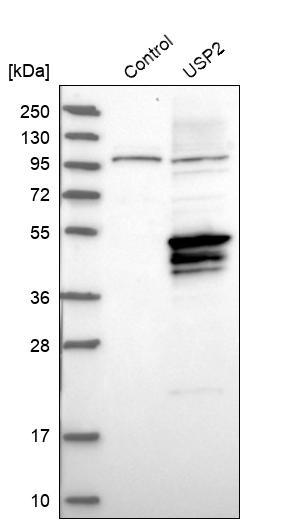
- Main image
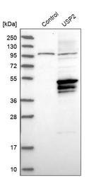
- Experimental details
- Western blot analysis in control (vector only transfected HEK293T lysate) and USP2 over-expression lysate (Co-expressed with a C-terminal myc-DDK tag (~3.1 kDa) in mammalian HEK293T cells, LY403527).
Supportive validation
- Submitted by
- Atlas Antibodies (provider)
- Main image

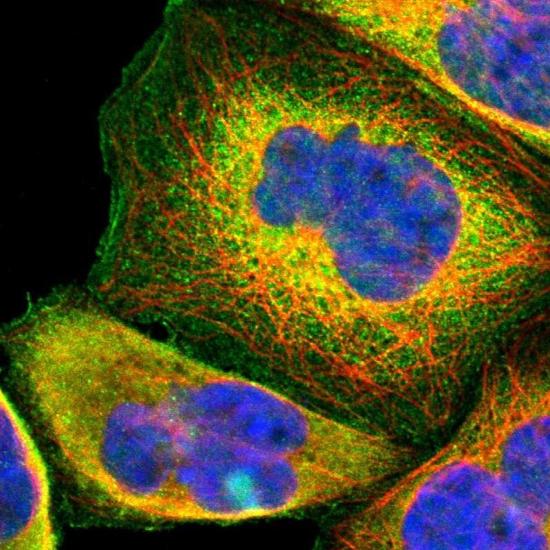
- Experimental details
- Immunofluorescent staining of human cell line A-431 shows localization to plasma membrane & cytosol.
- Sample type
- HUMAN
- Submitted by
- Atlas Antibodies (provider)
- Main image

- Experimental details
- Immunofluorescent staining of human cell line A-431 shows localization to plasma membrane & cytosol.
- Sample type
- Human
- Protocol
- Protocol

Supportive validation
- Submitted by
- Atlas Antibodies (provider)
- Main image

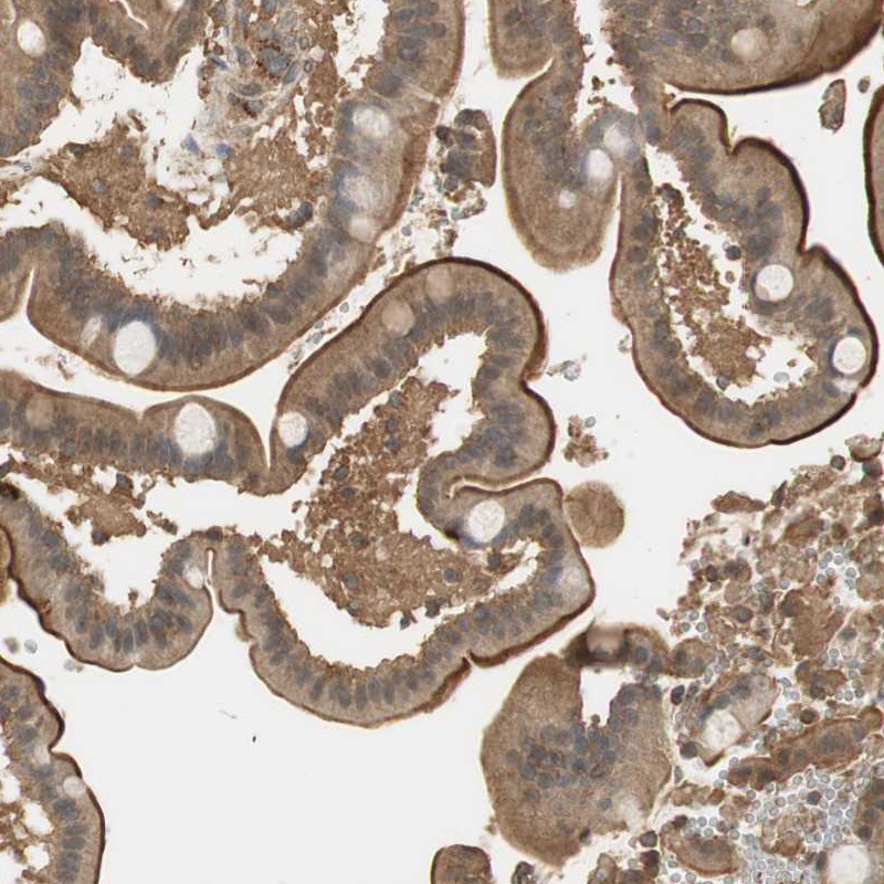
- Experimental details
- Immunohistochemical staining of human duodenum shows strong luminal membranous and cytoplasmic positivity in glandular cells.
- Sample type
- HUMAN
- Submitted by
- Atlas Antibodies (provider)
- Main image

- Experimental details
- Immunohistochemical staining of human duodenum shows strong luminal membranous and cytoplasmic positivity in glandular cells.
- Sample type
- Human
- Protocol
- Protocol
