 Explore
 Validate
 Learn
Learn Western blot
 Immunoprecipitation
Antibody data
- Antibody Data
- Antigen structure
- References [0]
- Comments [0]
- Validations
- Western blot [4]
- Immunocytochemistry [2]
- Immunohistochemistry [5]
- Other assay [1]
Submit
Validation data
Reference
Comment
Report error
- Product number
- 10881-1-AP - Provider product page

- Provider
- Invitrogen Antibodies
- Product name
- PTGES2 Polyclonal Antibody
- Antibody type
- Polyclonal
- Antigen
- Other
- Description
- Immunogen sequence: MDPAARVVR ALWPGGCALA WRLGGRPQPL LPTQSRAGFA GAAGGPSPVA AARKGSPRLL GAAALALGGA LGLYHTARWH LRAQDLHAER SAAQLSLSSR LQLTLYQYKT CPFCSKVRAF LDFHALPYQV VEVNPVRRAE IKFSSYRKVP ILVAQEGESS QQLNDSSVII SALKTYLVSG QPLEEIITYY PAMKAVNEQG KEVTEFGNKY WLMLNEKEAQ QVYGGKEART EEMKWRQWAD DWLVHLISPN VYRTPTEALA SFDYIVREGK FGAVEGAVAK YMGAAAMYLI SKRLKSRHRL QDNVREDLYE AADKWVAAVG KDRPFMGGQK PNLADLAVYG VLRVMEGLDA FDDLMQHTHI QPWYLRVERA ITEASPAH (1-377 aa encoded by BC011613)
- Reactivity
- Human, Mouse, Rat
- Host
- Rabbit
- Isotype
- IgG
- Vial size
- 150 µL
- Concentration
- 0.13 mg/mL
- Storage
- -20°C
No comments: Submit comment
Supportive validation
- Submitted by
- Invitrogen Antibodies (provider)
- Main image
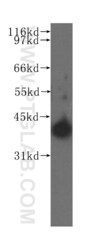
- Experimental details
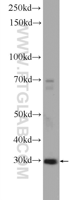
- Human stomach tissue were subjected to SDS PAGE followed by western blot with 10881-1-AP (PTGES2 antibody) at dilution of 1:400 incubated at room temperature for 1.5 hours.

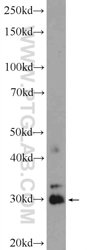
- Submitted by
- Invitrogen Antibodies (provider)
- Main image
- Experimental details
- HeLa cells were subjected to SDS PAGE followed by western blot with 10881-1-AP ( PTGES2 Antibody) at dilution of 1:600 incubated at room temperature for 1.5 hours.
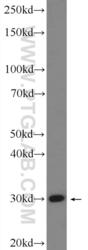
- Submitted by
- Invitrogen Antibodies (provider)
- Main image

- Experimental details
- Mouse heart tissue were subjected to SDS PAGE followed by western blot with 10881-1-AP (PTGES2 Antibody) at dilution of 1:600 incubated at room temperature for 1.5 hours.
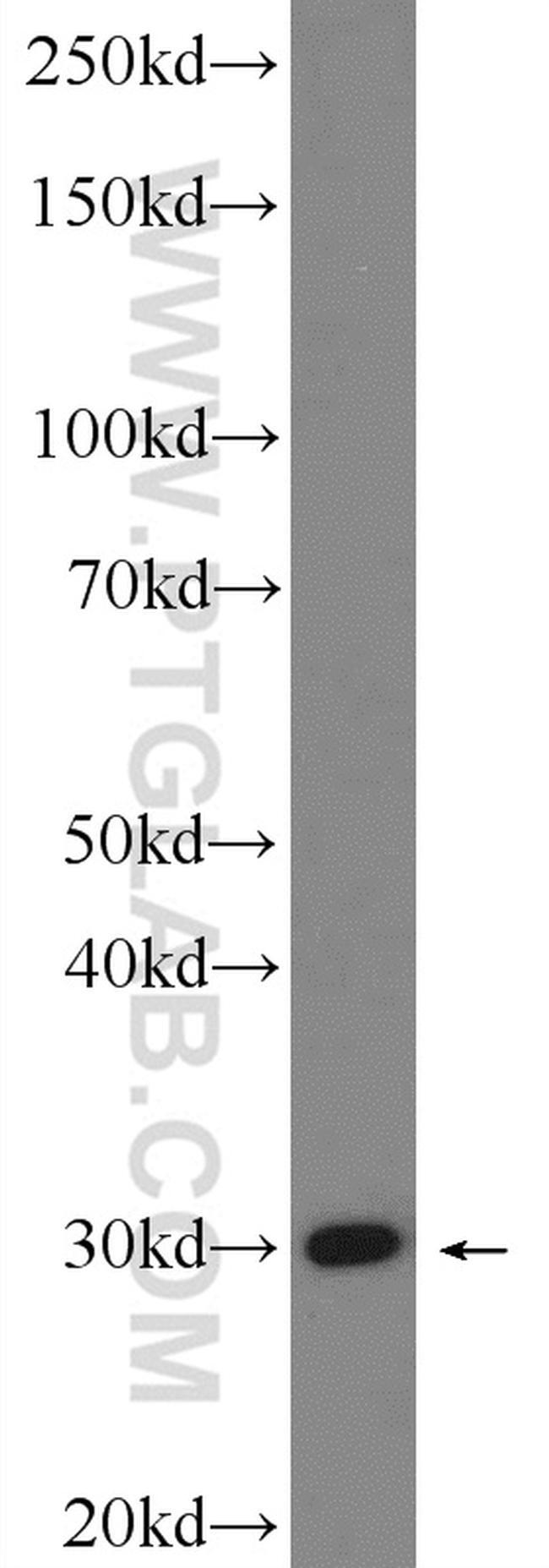
- Submitted by
- Invitrogen Antibodies (provider)
- Main image

- Experimental details
- HEK-293 cells were subjected to SDS PAGE followed by western blot with 10881-1-AP (PTGES2 Antibody) at dilution of 1:600 incubated at room temperature for 1.5 hours.
Supportive validation
- Submitted by
- Invitrogen Antibodies (provider)
- Main image
- Experimental details
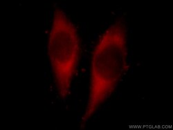
- Immunofluorescent analysis of Hela cells, using PTGES2 antibody 10881-1-AP at 1:25 dilution and Rhodamine-labeled goat anti-rabbit IGG (red).

- Submitted by
- Invitrogen Antibodies (provider)
- Main image

- Experimental details
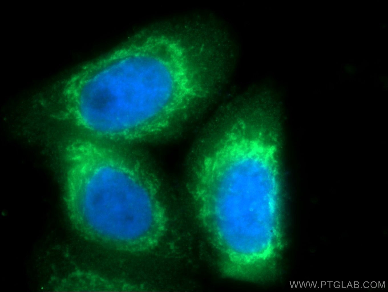
- Immunofluorescent analysis of HepG2 cells using 10881-1-AP (PTGES2 antibody) at dilution of 1:25 and Alexa Fluor 488-conjugated AffiniPure Goat Anti-Rabbit IgG (H+L).
Supportive validation
- Submitted by
- Invitrogen Antibodies (provider)
- Main image

- Experimental details
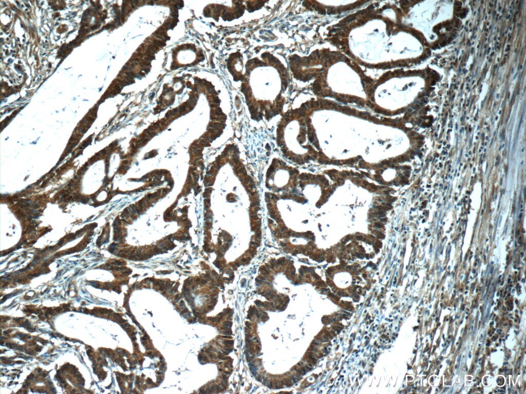
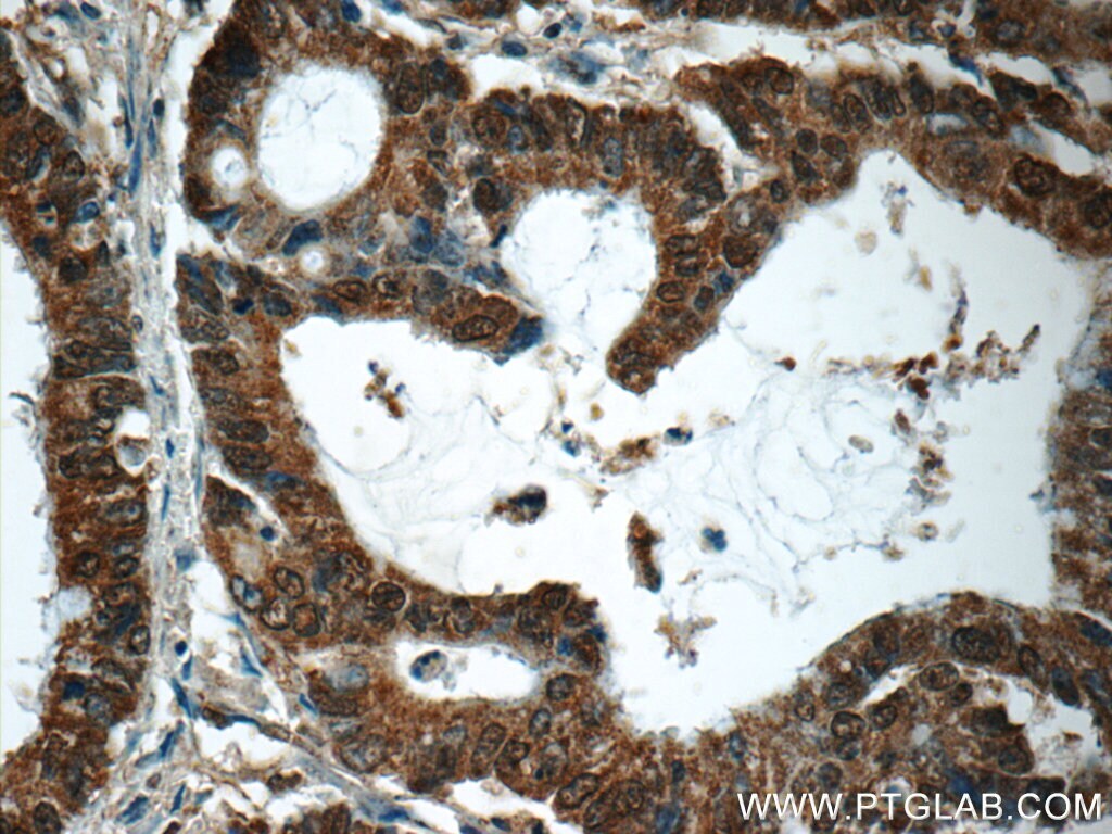
- Immunohistochemistry of paraffin-embedded human colon cancer tissue slide using 10881-1-AP (PTGES2 Antibody) at dilution of 1:50 (under 10x lens).
- Submitted by
- Invitrogen Antibodies (provider)
- Main image

- Experimental details
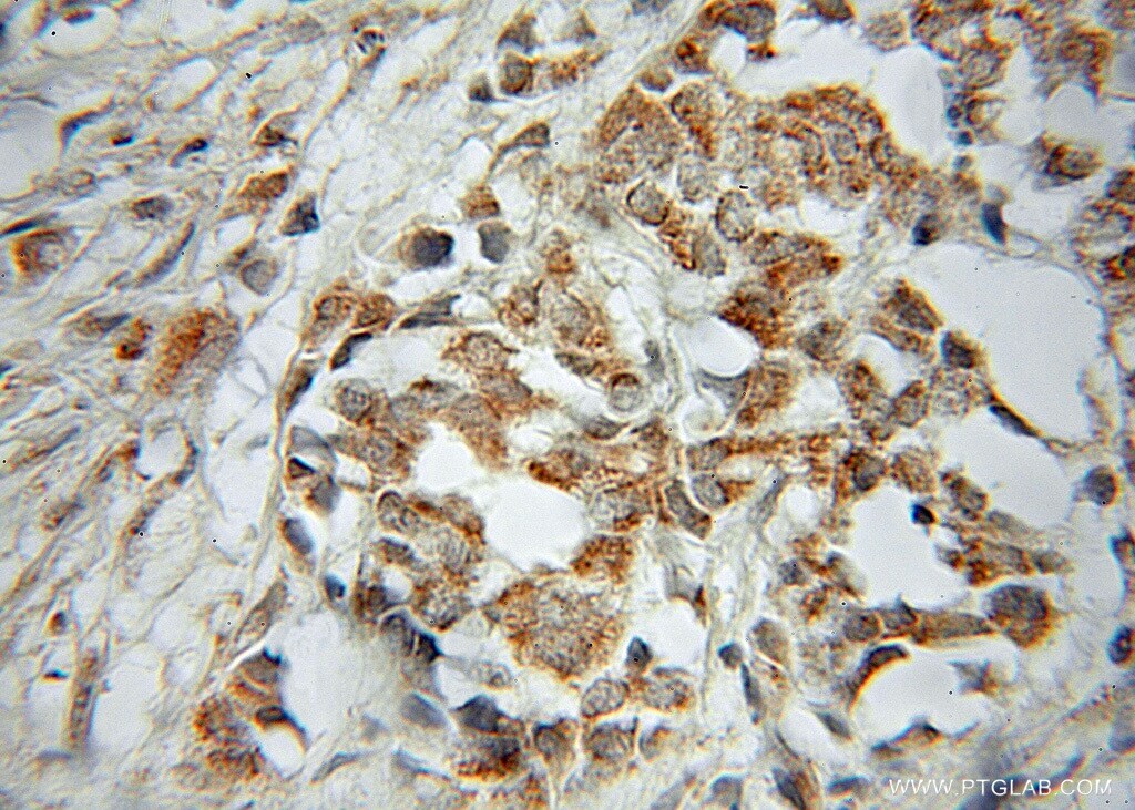
- Immunohistochemistry of paraffin-embedded human colon cancer tissue slide using 10881-1-AP (PTGES2 Antibody) at dilution of 1:50 (under 40x lens).
- Submitted by
- Invitrogen Antibodies (provider)
- Main image
- Experimental details
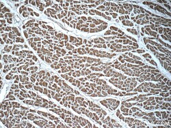
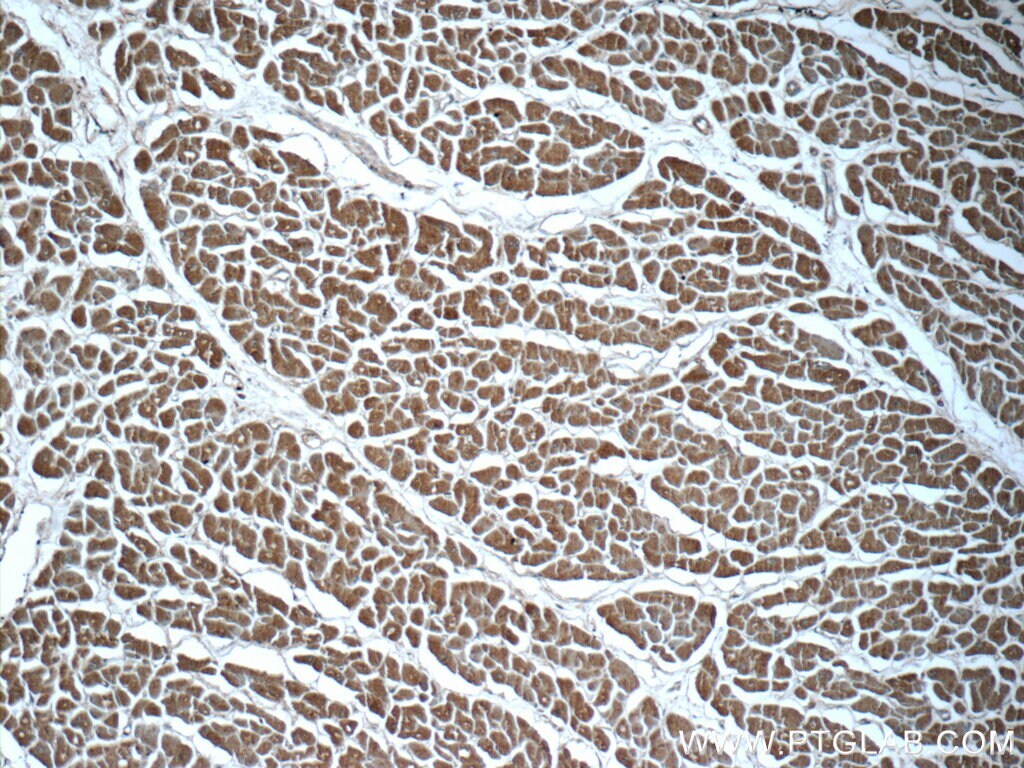
- Immunohistochemistry of paraffin-embedded human heart tissue slide using 10881-1-AP (PTGES2 Antibody) at dilution of 1:50 (under 10x lens).
- Submitted by
- Invitrogen Antibodies (provider)
- Main image

- Experimental details
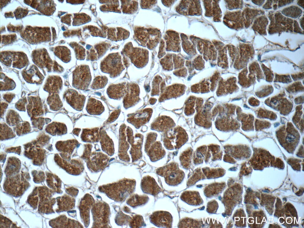
- Immunohistochemistry of paraffin-embedded human heart tissue slide using 10881-1-AP (PTGES2 Antibody) at dilution of 1:50 (under 40x lens).
- Submitted by
- Invitrogen Antibodies (provider)
- Main image

- Experimental details
- Immunohistochemistry of paraffin-embedded human breast cancer using 10881-1-AP (PTGES2 antibody) at dilution of 1:50 (under 40x lens).
Supportive validation
- Submitted by
- Invitrogen Antibodies (provider)
- Main image
- Experimental details
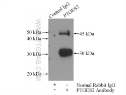
- IP result of anti-PTGES2 (IP:10881-1-AP, 4ug; Detection:10881-1-AP 1:500) with mouse heart tissue lysate 4000ug.
