 Explore
 Validate
 Learn
Learn Western blot
Antibody data
- Antibody Data
- Antigen structure
- References [0]
- Comments [0]
- Validations
- Western blot [2]
- Immunocytochemistry [1]
Submit
Validation data
Reference
Comment
Report error
- Product number
- PA5-109948 - Provider product page

- Provider
- Invitrogen Antibodies
- Product name
- MBTPS1 Polyclonal Antibody
- Antibody type
- Polyclonal
- Antigen
- Recombinant full-length protein
- Reactivity
- Human, Mouse
- Host
- Rabbit
- Isotype
- IgG
- Vial size
- 100 µL
- Concentration
- 3.46 mg/mL
- Storage
- -20° C, Avoid Freeze/Thaw Cycles
No comments: Submit comment
Supportive validation
- Submitted by
- Invitrogen Antibodies (provider)
- Main image

- Experimental details
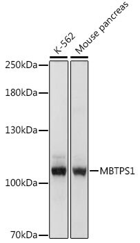
- Western blot analysis of MBTPS1 in K-562, mouse pancreas. Samples were incubated in polyclonal MBTPS1 antibody (Product # PA5-109948) using a dilution of 1:1000, followed by HRP Goat Anti-Rabbit IgG (H+L) at a dilution of 1:10000.
- Submitted by
- Invitrogen Antibodies (provider)
- Main image
- Experimental details
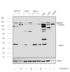
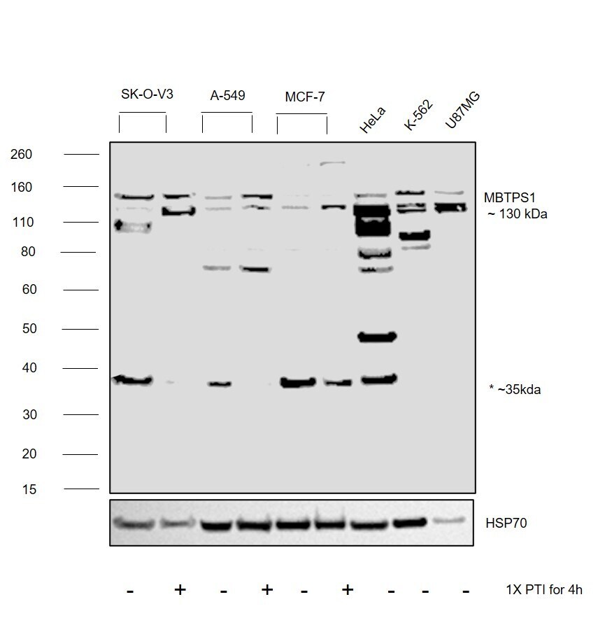
- Western blot was performed using Anti-MBTPS1 Polyclonal Antibody (Product # PA5-109948) and a ~130 kDa band corresponding to Membrane-bound transcription factor site-1 protease was observed across cell lines tested . Membrane enriched extracts (50 µg lysate) of SK-O-V3 (Lane 1), SK-O-V3 treated with PTI (Lane 2), A549 (Lane 3), A549 treated with PTI (Lane 4), MCF7 (Lane 5), MCF7 treated with PTI (Lane 6), HeLa (Lane 7), K-562 (Lane 8), u-87MG (Lane 9) were electrophoresed using NuPAGE™ 4-12% Bis-Tris Protein Gel (Product # NP0321BOX). Resolved proteins were then transferred onto a nitrocellulose membrane (Product # IB23001) by iBlot® 2 Dry Blotting System (Product # IB21001). The blot was probed with the primary antibody (1:1000) and detected by chemiluminescence with Goat anti-Rabbit IgG (H+L) Superclonal™ Recombinant Secondary Antibody, HRP (Product # A27036,1:20000) using the iBright FL 1000 (Product # A32752). Chemiluminescent detection was performed using SuperSignal™ West Pico PLUS Chemiluminescent Substrate (Product # 34580). A non specific band was observed around 35 kDa. Increased expression was observed in cell lines post PTI treatment.
Supportive validation
- Submitted by
- Invitrogen Antibodies (provider)
- Main image
- Experimental details
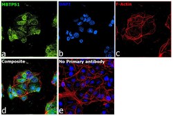
- Immunofluorescence analysis of Membrane-bound transcription factor site-1 protease was performed using 70% confluent log phase MCF7 cells. The cells were fixed with 4% paraformaldehyde for 10 minutes, permeabilized with 0.1% Triton™ X-100 for 15 minutes, and blocked with 2% BSA for 45 minutes at room temperature. The cells were labeled with MBTPS1 Polyclonal Antibody (Product # PA5-109948) at 1:100 in 0.1% BSA, incubated at 4 degree celsius overnight and then labeled with Donkey anti-Rabbit IgG (H+L) Highly Cross-Adsorbed Secondary Antibody, Alexa Fluor Plus 488 (Product # A32790), (1:2000), for 45 minutes at room temperature (Panel a: Green). Nuclei (Panel b:Blue) were stained with ProLong™ Diamond Antifade Mountant with DAPI (Product # P36962). F-actin (Panel c: Red) was stained with Rhodamine Phalloidin (Product # R415, 1:300). Panel d represents the merged image showing endoplasmic reticulum and golgi apparatus localization. Panel e represents control cells with no primary antibody to assess background. The images were captured at 60X magnification.
