 Explore
 Validate
 Learn
Learn Western blot
Antibody data
- Antibody Data
- Antigen structure
- References [2]
- Comments [0]
- Validations
- Western blot [2]
- Immunocytochemistry [1]
- Immunohistochemistry [2]
- Flow cytometry [1]
Submit
Validation data
Reference
Comment
Report error
- Product number
- 700180 - Provider product page

- Provider
- Invitrogen Antibodies
- Product name
- PSME3 Recombinant Rabbit Monoclonal Antibody (20H9L19)
- Antibody type
- Monoclonal
- Antigen
- Synthetic peptide
- Description
- This antibody is predicted to react with canine, chicken, equine, orangutan, Xenopus and zebrafish based on sequence homology.
- Antibody clone number
- 20H9L19
- Concentration
- 0.5 mg/mL
Submitted references Site-specific O-GlcNAcylation of Psme3 maintains mouse stem cell pluripotency by impairing P-body homeostasis.
Mutant p53 (p53-R248Q) functions as an oncogene in promoting endometrial cancer by up-regulating REGγ.
Pecori F, Kondo N, Ogura C, Miura T, Kume M, Minamijima Y, Yamamoto K, Nishihara S
Cell reports 2021 Jul 13;36(2):109361
Cell reports 2021 Jul 13;36(2):109361
Mutant p53 (p53-R248Q) functions as an oncogene in promoting endometrial cancer by up-regulating REGγ.
Wang H, Bao W, Jiang F, Che Q, Chen Z, Wang F, Tong H, Dai C, He X, Liao Y, Liu B, Sun J, Wan X
Cancer letters 2015 May 1;360(2):269-79
Cancer letters 2015 May 1;360(2):269-79
No comments: Submit comment
Supportive validation
- Submitted by
- Invitrogen Antibodies (provider)
- Main image
- Experimental details
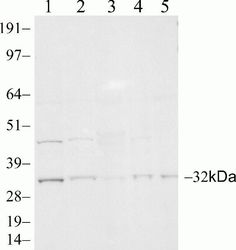
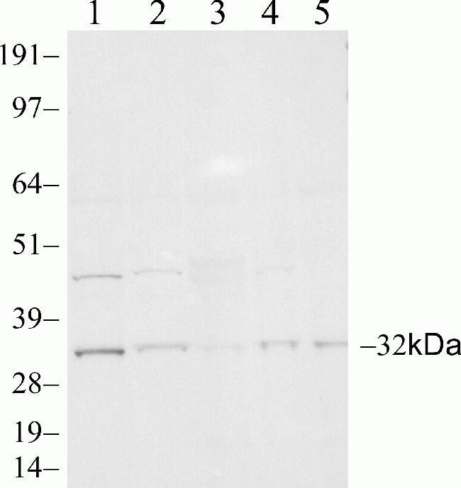
- Western blot analysis of PSME3 in Jurkat (lane 1), HeLa (lane 2), rat liver (lane 3), PC12 (lane 4), and mouse liver (lane 5) using a PSME3 recombinant rabbit monoclonal antibody (Product # 700180) at a dilution of 1 µg/mL.
- Submitted by
- Invitrogen Antibodies (provider)
- Main image
- Experimental details
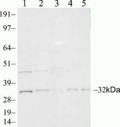
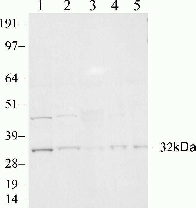
- Western blot analysis of PSME3 in Jurkat (lane 1), HeLa (lane 2), rat liver (lane 3), PC12 (lane 4), and mouse liver (lane 5) using a PSME3 recombinant rabbit monoclonal antibody (Product # 700180) at a dilution of 1 µg/mL.
Supportive validation
- Submitted by
- Invitrogen Antibodies (provider)
- Main image
- Experimental details
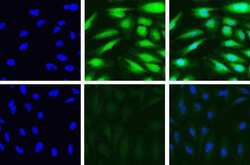
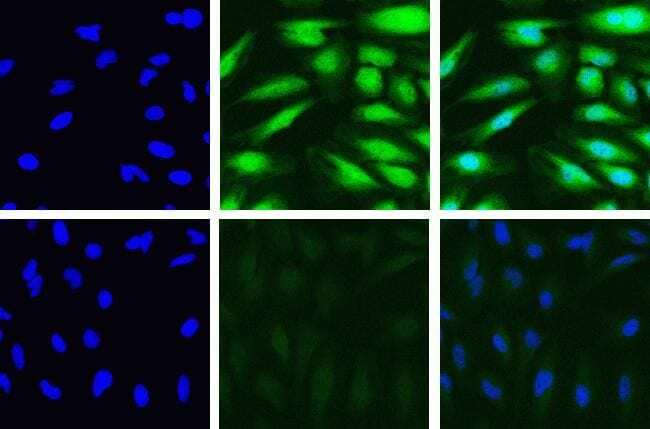
- Immunofluorescent analysis of PSME3 in HeLa cells using a PSME3 recombinant rabbit monoclonal antibody (Product # 700180) at a dilution of 5 µg/mL in the absence of peptide (top) and presence of immunogenic peptide (bottom), followed by detection using an Alexa Fluor 488-conjugated goat anti-rabbit secondary antibody at a dilution of 1:1000. Actin was stained with Alexa Fluor 568 Phalloidin (Product # A12380). Hoechst only (blue, left), AF488 signal only (green, middle) and composite image with Phalloidin (right).
Supportive validation
- Submitted by
- Invitrogen Antibodies (provider)
- Main image
- Experimental details
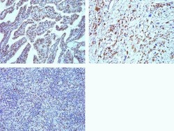
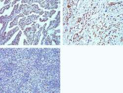
- Immunohistochemistry analysis of PSME3 in formalin-fixed, paraffin-embedded human thyroid (top left) and lung carcimona (top right) and Hodgkin lymphoma (bottom left) using a PSME3 monoclonal antibody (Product # 700180) at a dilution of 5 µg/mL. Staining was visualized using DAB and images were taken at a magnification of 20x. Results show strong nuclear staining in tumor cells.

- Submitted by
- Invitrogen Antibodies (provider)
- Main image
- Experimental details
- Immunohistochemistry analysis of PSME3 in formalin-fixed, paraffin-embedded human thyroid (top left) and lung carcimona (top right) and Hodgkin lymphoma (bottom left) using a PSME3 monoclonal antibody (Product # 700180) at a dilution of 5 µg/mL. Staining was visualized using DAB and images were taken at a magnification of 20x. Results show strong nuclear staining in tumor cells.

Supportive validation
- Submitted by
- Invitrogen Antibodies (provider)
- Main image
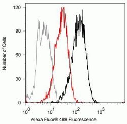
- Experimental details
- Flow cytometry analysis of PSME3 in Jurkat cells using a PSME3 recombinant rabbit monoclonal antibody (Product # 700180) at a dilution of 2ug. Cells were fixed and permeabilized using FIX & PERM (Product # GAS004) reagent, and detection was performed using an Alexa Fluor 488 goat anti-rabbit IgG (black) compared to a control without primary antibody (gray). Pre-incubation with the immunogenic peptide decreased the signal (red).
