 Explore
 Validate
 Learn
Learn Western blot
Antibody data
- Antibody Data
- Antigen structure
- References [1]
- Comments [0]
- Validations
- Western blot [3]
- Immunocytochemistry [2]
- Immunohistochemistry [2]
- Other assay [3]
Submit
Validation data
Reference
Comment
Report error
- Product number
- PA5-21789 - Provider product page

- Provider
- Invitrogen Antibodies
- Product name
- PSME3 Polyclonal Antibody
- Antibody type
- Polyclonal
- Antigen
- Recombinant protein fragment
- Description
- Recommended positive controls: 293T, A549, H1299, HCT116, MCF-7, NIH-3T3, JC, BCL-1, PC-12.
- Concentration
- 0.65 mg/mL
Submitted references Identification of a BRAF/PA28γ/MEK1 signaling axis and its role in epithelial-mesenchymal transition in oral submucous fibrosis.
Xie C, Li Z, Hua Y, Sun S, Zhong L, Chen Q, Feng H, Ji N, Li T, Zhou X, Zeng X, Tang Z, Sun C, Li J, Chen Q
Cell death & disease 2022 Aug 12;13(8):701
Cell death & disease 2022 Aug 12;13(8):701
No comments: Submit comment
Supportive validation
- Submitted by
- Invitrogen Antibodies (provider)
- Main image
- Experimental details
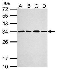
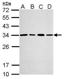
- Western Blot using PSME3 Polyclonal Antibody (Product # PA5-21789). Sample (30 µg of whole cell lysate). Lane A: A549. Lane B: H1299. Lane C: HCT116. Lane D: MCF-7. 12% SDS PAGE. PSME3 Polyclonal Antibody (Product # PA5-21789) diluted at 1:2,000. The HRP-conjugated anti-rabbit IgG antibody was used to detect the primary antibody.
- Submitted by
- Invitrogen Antibodies (provider)
- Main image
- Experimental details
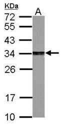
- Western Blot using PSME3 Polyclonal Antibody (Product # PA5-21789). Sample (30 µg of whole cell lysate). Lane A: PC-12. 12% SDS PAGE. PSME3 Polyclonal Antibody (Product # PA5-21789) diluted at 1:2,000. The HRP-conjugated anti-rabbit IgG antibody was used to detect the primary antibody.

- Submitted by
- Invitrogen Antibodies (provider)
- Main image

- Experimental details
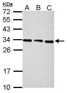
- Western Blot using PSME3 Polyclonal Antibody (Product # PA5-21789). Sample (30 µg of whole cell lysate). Lane A: NIH-3T3. Lane B: JC. Lane C: BCL-1. 12% SDS PAGE. PSME3 Polyclonal Antibody (Product # PA5-21789) diluted at 1:2,000. The HRP-conjugated anti-rabbit IgG antibody was used to detect the primary antibody.
Supportive validation
- Submitted by
- Invitrogen Antibodies (provider)
- Main image
- Experimental details
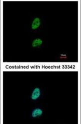
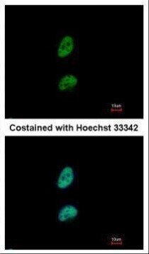
- Immunofluorescent analysis of PSME3 in paraformaldehyde-fixed HeLa cells using a PSME3 polyclonal antibody (Product # PA5-21789) at a 1:500 dilution.

- Submitted by
- Invitrogen Antibodies (provider)
- Main image

- Experimental details
- Immunofluorescence analysis of paraformaldehyde-fixed HeLa, using PSME3 (Product # PA5-21789) antibody at 1:500 dilution.
Supportive validation
- Submitted by
- Invitrogen Antibodies (provider)
- Main image
- Experimental details
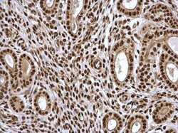
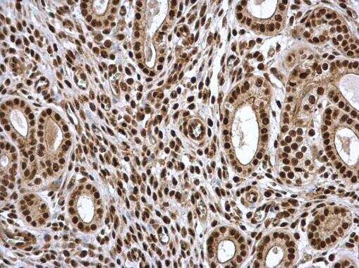
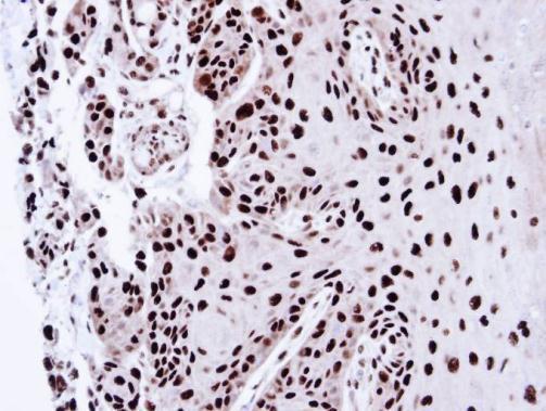
- Immunohistochemistry (Paraffin) analysis of PSME3 was performed in paraffin-embedded mouse cervix tissue using PSME3 Polyclonal Antibody (Product # PA5-21789) at a dilution of 1:500.
- Submitted by
- Invitrogen Antibodies (provider)
- Main image
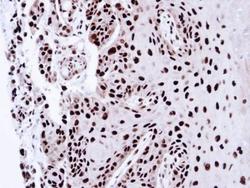
- Experimental details
- Immunohistochemical analysis of paraffin-embedded CA922 xenograft, using PSME3 (Product # PA5-21789) antibody at 1:100 dilution. Antigen Retrieval: Citrate buffer, pH 6.0, 15 min.
Supportive validation
- Submitted by
- Invitrogen Antibodies (provider)
- Main image
- Experimental details
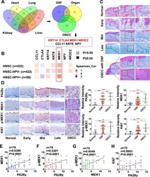
- The expression of PA28gamma in OSF epithelial tissue was positively correlated with both MEK1 and phosphorylated MEK1. A Obtaining OSF malignant transformation genes through database search (GeneCards) and bioinformatics analysis. A total of 709 organ fibrosis genes were associated with pulmonary fibrosis (2349), liver fibrosis (1778), renal fibrosis (1792) and myocardial fibrosis (883). A Venn diagram yielded 7 genes associated with organ fibrosis (709), OSF (181) and OSCC (1693). Among them, 4 upregulated genes (KRT14, CTLA4, MEK1, and NR3C2) and 3 downregulated genes (CCL11, KRT8, and NYP) were obtained. B Correlation analysis showed that the MEK1 gene positively correlated with PA28gamma. C Masson's trichrome staining for identifying different stages of OSF phenotype, including early, mid, late, and OSCC with OSF. Scale bar: left panels, 500 mum; mid panels, 200 mum; right panels, 100 mum. D IHC staining and statistical analysis showed the expression of PA28gamma, MEK1, p-MEK1, and Ki67 in normal ( n = 10), OSF ( n = 52), and OSCC with OSF ( n = 18) tissues. Based on Student's t test (* p < 0.05, ** p < 0.01, *** p < 0.001, **** p < 0.000). Scale bar: 200 mum. E - H Correlation analysis of 4 molecules in the normal, OSF and OSCC groups.

- Submitted by
- Invitrogen Antibodies (provider)
- Main image
- Experimental details
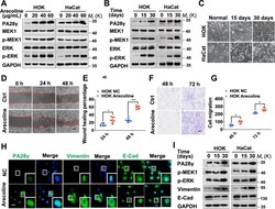
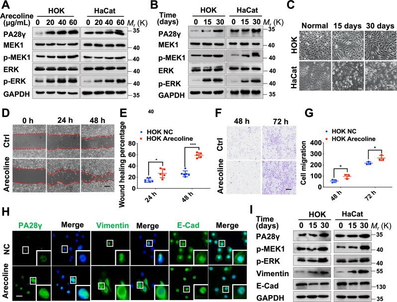
- Arecoline can upregulate the expression levels of PA28gamma and phosphorylated MEK1 in epithelial cells, which induce EMT. Western blot assays showed that arecoline upregulated the expression levels of PA28gamma, p-MEK1, and p-ERK in an adosage-dependent ( A ) and time-dependent manner ( B ). C After 30 days of treatment of epithelial cells with low concentrations of arecoline (10 mug/mL), the morphology of epithelial cells transformed to a mesenchymal phenotype. Scale bar: 25 mum. Wound healing ( D , E ) and transwell assays F , G showed that the healing ability and migration ability of epithelial cells treated with arecoline were upregulated. Scale bar: 100 mum. Data represent the means +- s.d. of three independent experiments. Statistical analysis was performed using Student's t test (* p < 0.05, *** p < 0.001). H Representative results for immunostaining of PA28gamma, Vimentin, and E-cadherin in the normal control group and the arecoline-treated group. Scale bar: 50 mum. I Western blot assays showed the expression levels of Vimentin and E-cadherin in arecoline-treated epithelial cells.
- Submitted by
- Invitrogen Antibodies (provider)
- Main image
- Experimental details
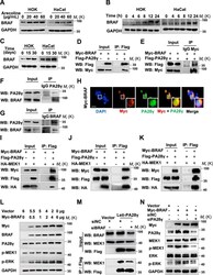
- Fig. 5 BRAF can bind to PA28gamma and stabilize its protein level. A - C Western blot assays showed that the expression levels of BRAF were upregulated in a dosage-dependent and time-dependent manner. D , E A coimmunoprecipitation assay showed that exogenous Myc-BRAF and Flag-PA28gamma bind to each other in 293T cells. F , G Coimmunoprecipitation experiments showed that endogenous BRAF and PA28gamma interact with each other in epithelial cells. H After transfecting the Myc-BRAF plasmid into 293T cells, coimmunostaining showed that Myc-BRAF (red) colocalized with PA28gamma (green). Scale bar: 50 mum. I Immunoprecipitation assays showed that exogenous BRAF and MEK could simultaneously bind to PA28gamma. Immunoprecipitation of PA28gamma was performed with the Flag antibody. J Exogenous BRAF and PA28gamma were detected in immunoprecipitates performed with the HA antibody. K Exogenous MEK1 and PA28gamma can be detected in immunoprecipitates performed with the Myc antibody. L Transfection of 293T cells with the Myc-BRAF plasmid in a dose-dependent manner. Western blot assays showed that the expression levels of PA28gamma, p-MEK1, and p-ERK were significantly upregulated. M Immunoprecipitation shows that silencing BRAF expression reduces MEK1 binding to PA28gamma. N Western blot assays showed that knocking down PA28gamma inhibited BRAF-dependent expression of p-MEK1 and p-ERK.
