 Explore
 Validate
 Learn
Learn Western blot
Antibody data
- Antibody Data
- Antigen structure
- References [1]
- Comments [0]
- Validations
- Western blot [2]
- Immunocytochemistry [1]
- Chromatin Immunoprecipitation [1]
Submit
Validation data
Reference
Comment
Report error
- Product number
- 703747 - Provider product page

- Provider
- Invitrogen Antibodies
- Product name
- ZNF207 Recombinant Rabbit Monoclonal Antibody (7H6L2)
- Antibody type
- Monoclonal
- Antigen
- Other
- Description
- This antibody is predicted to react with Bovine.
- Antibody clone number
- 7H6L2
- Concentration
- 0.5 mg/mL
Submitted references System analysis based on the cancer-immunity cycle identifies ZNF207 as a novel immunotherapy target for hepatocellular carcinoma.
Wang X, Zhou T, Chen X, Wang Y, Ding Y, Tu H, Gao S, Wang H, Tang X, Yang Y
Journal for immunotherapy of cancer 2022 Mar;10(3)
Journal for immunotherapy of cancer 2022 Mar;10(3)
No comments: Submit comment
Supportive validation
- Submitted by
- Invitrogen Antibodies (provider)
- Main image

- Experimental details
- Western blot was performed using Anti-ZNF207 Recombinant Rabbit Monoclonal Antibody (Product # 703747) and a 58 kDa band corresponding to ZNF207 was observed across cell lines and tissue tested. Modified Whole cell extract (1% SDS) (30 µg lysate) of HeLa (Lane 1), U-2 OS (Lane 2), HCT 116 (Lane 3), A549 (Lane 4), NIH/3T3 (Lane 5) and tissue extract of Mouse Lung (Lane 6) were electrophoresed using Novex® NuPAGE™ 4-12% Bis-Tris Protein Gel (Product # NP0321BOX). Resolved proteins were then transferred onto a nitrocellulose membrane (Product # IB23001) by iBlot® 2 Dry Blotting System (Product # IB21001). The blot was probed with the primary antibody (1:2000 dilution) and detected by chemiluminescence using Goat anti-Rabbit IgG (H+L) Superclonal™ Secondary Antibody, HRP conjugate (Product # A27036) at a 1:5000 dilution. Chemiluminescent detection was performed using Novex® ECL Chemiluminescent Substrate Reagent Kit (Product # WP20005).

- Submitted by
- Invitrogen Antibodies (provider)
- Main image

- Experimental details
- Knockdown of ZNF207 was achieved by transfecting HeLa cells with ZNF207 specific siRNA (Silencer® select Product # S15278, Product # s15280). Western blot analysis (Fig a) was performed using modified whole cell extracts (1% SDS) from ZNF207 knockdown cells (Lane 3), non-specific scrambled siRNA transfected cells (Lane 2) and untransfected cells (Lane 1). The blot was probed with Anti-ANF207 Recombinant Rabbit Monoclonal Antibody (Product # 703747) at a 1:2000 dilution and Goat anti-Rabbit IgG (H+L) Superclonal™ Secondary Antibody, HRP conjugate (Product # A27036) (0.25 µg/mL, 1:4000 dilution). Densitometric analysis of this western blot is shown in the histogram (Fig b). Loss of signal upon siRNA mediated knockdown confirms that the antibody is specific to ZNF207.

Supportive validation
- Submitted by
- Invitrogen Antibodies (provider)
- Main image
- Experimental details
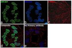
- For immunocytochemistry analysis, HeLa cells were fixed and permeabilized for detection of endogenous ZNF207 using Anti-ZNF207 Recombinant Rabbit Monoclonal Antibody (Product # 703747) at a 1:100 dilution and labeled with Goat anti-Rabbit IgG (H+L) Highly Cross-Adsorbed Secondary Antibody, Alexa Fluor Plus 488 conjugate (Product # A32731) at a 1:2000 dilution. Panel a) shows representative cells that were stained for detection and localization of ZNF207 protein (green), Panel b) is stained for nuclei (blue) using ProLong™ Diamond Antifade Mountant with DAPI (Product # P36962). Panel c) represents cytoskeletal F-actin staining using Rhodamine Phalloidin (Product # R415) at a 1:300 dilution. Panel d) is a composite image of Panels a, b and c clearly demonstrating nuclear localization of ZNF207. Panel e) represents control cells with no primary antibody to assess background. The images were captured at 60X magnification.

Supportive validation
- Submitted by
- Invitrogen Antibodies (provider)
- Main image

- Experimental details
- Chromatin Immunoprecipitation (ChIP) assay of endogenous ZNF207 protein using Anti-ZNF207 Antibody: ChIP was performed using 5 µg of Anti-ZNF207 Recombinant Rabbit Monoclonal Antibody (Product # 703747) on sheared chromatin from 2 million HeLa cells using the MAGnify ChIP System kit (Product # 49-2024). Normal Rabbit IgG was used as a negative IP control. The purified DNA was analyzed by qPCR using primers binding to the transcriptional start site of FGF2, Neurog3, Sall4, Nes and SAT2 satellite repeats. Data is presented as fold enrichment of the antibody signal versus the negative control IgG using the comparative CT method.
