Explore
 Validate
 Learn
Learn Western blot
Antibody data
- Antibody Data
- Antigen structure
- References [4]
- Comments [0]
- Validations
- Western blot [2]
- Immunoprecipitation [1]
- Immunohistochemistry [1]
Submit
Validation data
Reference
Comment
Report error
- Product number
- NB100-1704 - Provider product page

- Provider
- Novus Biologicals
- Proper citation
- Novus Cat#NB100-1704, RRID:AB_10000901
- Product name
- Rabbit Polyclonal BTF Antibody
- Antibody type
- Polyclonal
- Description
- Immunogen affinity purified.
- Reactivity
- Human, Mouse
- Host
- Rabbit
- Isotype
- IgG
- Vial size
- 0.1 ml
- Concentration
- 0.2 mg/ml
- Storage
- Store at 4C. Do not freeze.
Submitted references BclAF1 restriction factor is neutralized by proteasomal degradation and microRNA repression during human cytomegalovirus infection.
BCLAF1 is a radiation-induced H2AX-interacting partner involved in γH2AX-mediated regulation of apoptosis and DNA repair.
Tandem array-based expression screens identify host mRNA targets of virus-encoded microRNAs.
Protein kinase C delta induces transcription of the TP53 tumor suppressor gene by controlling death-promoting factor Btf in the apoptotic response to DNA damage.
Lee SH, Kalejta RF, Kerry J, Semmes OJ, O'Connor CM, Khan Z, Garcia BA, Shenk T, Murphy E
Proceedings of the National Academy of Sciences of the United States of America 2012 Jun 12;109(24):9575-80
Proceedings of the National Academy of Sciences of the United States of America 2012 Jun 12;109(24):9575-80
BCLAF1 is a radiation-induced H2AX-interacting partner involved in γH2AX-mediated regulation of apoptosis and DNA repair.
Lee YY, Yu YB, Gunawardena HP, Xie L, Chen X
Cell death & disease 2012 Jul 26;3:e359
Cell death & disease 2012 Jul 26;3:e359
Tandem array-based expression screens identify host mRNA targets of virus-encoded microRNAs.
Ziegelbauer JM, Sullivan CS, Ganem D
Nature genetics 2009 Jan;41(1):130-4
Nature genetics 2009 Jan;41(1):130-4
Protein kinase C delta induces transcription of the TP53 tumor suppressor gene by controlling death-promoting factor Btf in the apoptotic response to DNA damage.
Liu H, Lu ZG, Miki Y, Yoshida K
Molecular and cellular biology 2007 Dec;27(24):8480-91
Molecular and cellular biology 2007 Dec;27(24):8480-91
No comments: Submit comment
Supportive validation
- Submitted by
- Novus Biologicals (provider)
- Main image
- Experimental details
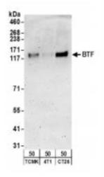
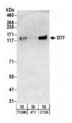
- Western Blot: BTF Antibody [NB100-1704] - : Whole cell lysate (50 ug) from TCMK-1, 4T1, and CT26.WT cells.
- Submitted by
- Novus Biologicals (provider)
- Main image

- Experimental details
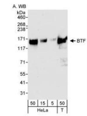
- Western Blot: BTF Antibody [NB100-1704] - Whole cell lysate from HeLa (5, 15, 50 ug loaded) and 293T cells (T; 50 ug) probed with anti-BTF antibody at 0.04 ug/ml.
Supportive validation
- Submitted by
- Novus Biologicals (provider)
- Main image

- Experimental details
- Immunoprecipitation: BTF Antibody [NB100-1704] - Whole cell lysate from HeLa (5, 15 and 50 ug for WB; 1 mg for IP, 20% of IP loaded) and 293T (T; 50 ug) cells.

Supportive validation
- Submitted by
- Novus Biologicals (provider)
- Main image
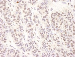
- Experimental details
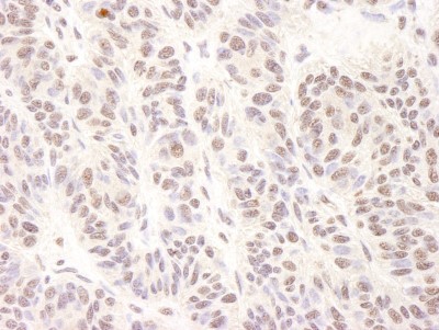
- Immunohistochemistry: BTF Antibody [NB100-1704] - Sample: FFPE section of human skin carcinoma. Antibody: Affinity purified rabbit anti-BTF used at a dilution of 1:1,000 (0.2ug/ml). Detection: DAB staining using Immunohistochemistry Accessory Kit.