 Explore
 Validate
 Learn
Learn Western blot
 Immunohistochemistry
Antibody data
- Antibody Data
- Antigen structure
- References [2]
- Comments [0]
- Validations
- Western blot [1]
- Immunocytochemistry [1]
Submit
Validation data
Reference
Comment
Report error
- Product number
- ABIN1580458 - Provider product page

- Provider
- antibodies-online
- Product name
- anti-Rhodopsin (RHO) antibody
- Antibody type
- Monoclonal
- Antigen
- Other
- Description
- affinity purified antibody
- Reactivity
- Human, Mouse, Rat, Bovine, Porcine
- Host
- Mouse
- Isotype
- IgG
- Antibody clone number
- B630
- Vial size
- 100 μL
- Concentration
- 1 mg/mL
- Storage
- Store at 4°C short term or -20°C long term.
- Handling
- Avoid repeated freezing and thawing.
Submitted references Photoreceptor membrane proteins, phototransduction, and retinal degenerative diseases. The Friedenwald Lecture.
Phototransduction mechanism in retinal rods and cones. The Friedenwald Lecture.
Molday RS
Investigative ophthalmology & visual science 1998 Dec;39(13):2491-513
Investigative ophthalmology & visual science 1998 Dec;39(13):2491-513
Phototransduction mechanism in retinal rods and cones. The Friedenwald Lecture.
Yau KW
Investigative ophthalmology & visual science 1994 Jan;35(1):9-32
Investigative ophthalmology & visual science 1994 Jan;35(1):9-32
No comments: Submit comment
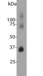
Supportive validation
- Submitted by
- antibodies-online (provider)
- Main image

- Experimental details
- Blot of bovine retinal extracts probed with ABIN1580458. The antibody stains a band corresponding to retinal rhodopsin at about 35 kDa. Bands about 70 kDa and 140 kDa are aggregated forms of rhodopsin. Note, due to the highly hydrophobic nature of rhodopsin, it important to avoid boiling samples containing this protein it in SDS-PAGE sample buffer, as this will result in even more extensive aggregation of the rhodopsin protein and appearance of more of this high molecular weight material.
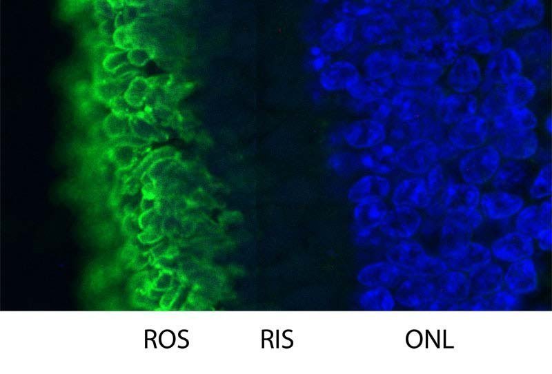
Supportive validation
- Submitted by
- antibodies-online (provider)
- Main image
- Experimental details
- High magnification confocal image of pig retinal section stained with ABIN1580458 (Green). Rhodopsin is most abundant in the rod outer segments (ROS) of retina, clearly localized in rod membranes. The rod inner segments (RIS) and rod nuclei in the outer nuclear layer (ONL) are also seen in this image. Nuclear DNA was stained with DAPI (blue).
