Antibody data
- Antibody Data
- Antigen structure
- References [2]
- Comments [0]
- Validations
- Western blot [5]
- Immunocytochemistry [1]
- Immunohistochemistry [1]
Submit
Validation data
Reference
Comment
Report error
- Product number
- GTX102595 - Provider product page
- Provider
- GeneTex
- Proper citation
- GeneTex Cat#GTX102595, RRID:AB_2036128
- Product name
- 53BP1 antibody [N1], N-term
- Antibody type
- Polyclonal
- Reactivity
- Human, Mouse
- Host
- Rabbit
Submitted references Genome Stability by DNA Polymerase β in Neural Progenitors Contributes to Neuronal Differentiation in Cortical Development.
Middle infrared radiation induces G2/M cell cycle arrest in A549 lung cancer cells.
Onishi K, Uyeda A, Shida M, Hirayama T, Yagi T, Yamamoto N, Sugo N
The Journal of neuroscience : the official journal of the Society for Neuroscience 2017 Aug 30;37(35):8444-8458
Middle infrared radiation induces G2/M cell cycle arrest in A549 lung cancer cells.
Chang HY, Shih MH, Huang HC, Tsai SR, Juan HF, Lee SC
PloS one 2013;8(1):e54117
No comments: Submit comment
Enhanced validation
Supportive validation
- Submitted by
- GeneTex (provider)
- Enhanced method
- Genetic validation
- Main image
- Experimental details
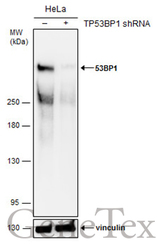
- Non-transfected (¡V) and transfected (+) HeLa whole cell extracts (50 ?g) were separated by 5% SDS-PAGE, and the membrane was blotted with 53BP1 antibody [N1], N-term (GTX102595) diluted at 1:500.
Supportive validation
- Submitted by
- GeneTex (provider)
- Main image
- Experimental details
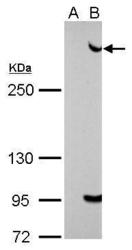
- 53BP1 antibody [N1], N-term detects TP53BP1 protein by Western blot analysis.A. 30 ug 293T whole cell lysate/extractB. 30 whole cell lysate/extract of human 53BP1-transfected 293T cells5 % SDS-PAGE53BP1 antibody [N1], N-term (GTX102595) dilution: 1:5000
- Validation comment
- WB
- Submitted by
- GeneTex (provider)
- Main image
- Experimental details
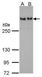
- 53BP1 antibody [N1], N-term detects TP53BP1 protein by Western blot analysis.A. 30 ?g 293T whole cell lysate/extractB. 30 ?g HeLa whole cell lysate/extract5 % SDS-PAGE53BP1 antibody [N1], N-term (GTX102595) dilution: 1:500
- Submitted by
- GeneTex (provider)
- Main image
- Experimental details
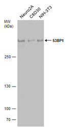
- Various whole cell extracts (30 £gg) were separated by 5% SDS-PAGE, and the membrane was blotted with 53BP1 antibody [N1], N-term (GTX102595) diluted at 1:500.
- Submitted by
- GeneTex (provider)
- Main image
- Experimental details
- Non-transfected (¡V) and transfected (+) HeLa whole cell extracts (50 ?g) were separated by 5% SDS-PAGE, and the membrane was blotted with 53BP1 antibody [N1], N-term (GTX102595) diluted at 1:500.
Supportive validation
- Submitted by
- GeneTex (provider)
- Main image
- Experimental details
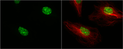
- 53BP1 antibody [N1], N-term detects 53BP1 protein at nucleus by immunofluorescent analysis.Sample: HeLa cells were fixed in 4% paraformaldehyde at RT for 15 min.Green: 53BP1 protein stained by 53BP1 antibody [N1], N-term (GTX102595) diluted at 1:500.Red: alpha Tubulin, a cytoskeleton marker, stained by alpha Tubulin antibody [B-5-1-2] (GTX11304) diluted at 1:10000.
Supportive validation
- Submitted by
- GeneTex (provider)
- Main image
- Experimental details
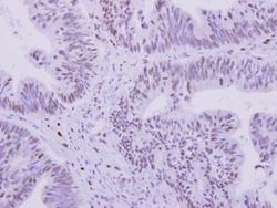
- 53BP1 antibody [N1], N-term detects 53BP1 protein at nucleus on human colon carcinoma by immunohistochemical analysis. Sample: Paraffin-embedded human colon carcinoma. 53BP1 antibody [N1], N-term (GTX102595) dilution: 1:250.