Antibody data
- Antibody Data
- Antigen structure
- References [0]
- Comments [0]
- Validations
- Western blot [1]
- Immunocytochemistry [2]
- Flow cytometry [1]
Submit
Validation data
Reference
Comment
Report error
- Product number
- TA309918 - Provider product page

- Provider
- OriGene
- Product name
- Rabbit Polyclonal 53BP1 Antibody
- Antibody type
- Polyclonal
- Description
- Rabbit Polyclonal 53BP1 Antibody
- Host
- Rabbit
- Conjugate
- Unconjugated
- Epitope
- TP53BP1
- Antibody clone number
- NULL
- Vial size
- 100 µl
- Concentration
- 1mg/ml
No comments: Submit comment
Supportive validation
- Submitted by
- OriGene (provider)
- Main image

- Experimental details
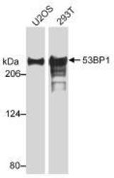
- Western Blot: 53BP1 Antibody - Detection of Human [deleted mouse] 53BP1 by Western Blot Sample: Whole cell lysate (20 ug/lane) from U2OS or 293T cells resolved on a 3 to 8% tris-acetate gel. Anti-53BP1 used at 0.5 ug/ml.
- Validation comment
- WB
Supportive validation
- Submitted by
- OriGene (provider)
- Main image

- Experimental details
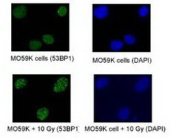
- Immunofluorescence: 53BP1 antibody - 53BP1 foci in proliferating MO59K cells; both normal and exposed to 10 Gy of IR and double-color immunofluorescence stained after 2 hours. Images captured in a Kodak digital image system on a Leica fluorescent microscope.
- Validation comment
- IF
- Submitted by
- OriGene (provider)
- Main image

- Experimental details
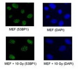
- immunofluorescence: 53BP1 amtibody - 53BP1 foci in proliferating MEFs; both normal and exposed to 10 Gy of IR and double-color immunofluorescence stained after 2 hours. Images captured in a Kodak digital image system on a Leica fluorescent microscope.
- Validation comment
- IF
Supportive validation
- Submitted by
- OriGene (provider)
- Main image

- Experimental details
- Flow Cytometry: 53BP1 Antibody - 1 million Jurkat cells were fixed, permeabilized, and stained with 1.5 ug/ml anti-53BP1 in a 150 ul reaction. Isotype control (black), anti-53BP1 (red).
- Validation comment
- FC
 Explore
 Validate
 Learn
Learn Western blot