 Explore
 Validate
 Learn
Learn Western blot
 ELISA
ELISAAntibody data
- Antibody Data
- Antigen structure
- References [9]
- Comments [0]
- Validations
- Western blot [9]
- ELISA [1]
- Immunocytochemistry [1]
- Immunoprecipitation [1]
Submit
Validation data
Reference
Comment
Report error
- Product number
- GTX118736 - Provider product page

- Provider
- GeneTex
- Proper citation
- GeneTex Cat#GTX118736, RRID:AB_10619853
- Product name
- TSG101 antibody
- Antibody type
- Polyclonal
- Reactivity
- Human, Mouse
- Host
- Rabbit
Submitted references Cancer-Specifically Re-Spliced TSG101 mRNA Promotes Invasion and Metastasis of Nasopharyngeal Carcinoma.
EIF3C-enhanced exosome secretion promotes angiogenesis and tumorigenesis of human hepatocellular carcinoma.
Cytosolic Genomic DNA functions as a Natural Antisense.
Transfer of Mammary Gland-forming Ability Between Mammary Basal Epithelial Cells and Mammary Luminal Cells via Extracellular Vesicles/Exosomes.
Analysis of Assembly and Budding of Lujo Virus.
The innate immune factor apolipoprotein L1 restricts HIV-1 infection.
Hrs- and CD63-dependent competing mechanisms make different sized endosomal intraluminal vesicles.
IDOL stimulates clathrin-independent endocytosis and multivesicular body-mediated lysosomal degradation of the low-density lipoprotein receptor.
Old world arenaviruses enter the host cell via the multivesicular body and depend on the endosomal sorting complex required for transport.
Chua HH, Kameyama T, Mayeda A, Yeh TH
International journal of molecular sciences 2019 Feb 12;20(3)
International journal of molecular sciences 2019 Feb 12;20(3)
EIF3C-enhanced exosome secretion promotes angiogenesis and tumorigenesis of human hepatocellular carcinoma.
Lee HY, Chen CK, Ho CM, Lee SS, Chang CY, Chen KJ, Jou YS
Oncotarget 2018 Mar 2;9(17):13193-13205
Oncotarget 2018 Mar 2;9(17):13193-13205
Cytosolic Genomic DNA functions as a Natural Antisense.
Asada K, Ito K, Yui D, Tagaya H, Yokota T
Scientific reports 2018 Jun 4;8(1):8551
Scientific reports 2018 Jun 4;8(1):8551
Transfer of Mammary Gland-forming Ability Between Mammary Basal Epithelial Cells and Mammary Luminal Cells via Extracellular Vesicles/Exosomes.
Lin MC, Chen SY, He PL, Luo WT, Li HJ
Journal of visualized experiments : JoVE 2017 Jun 3;(124)
Journal of visualized experiments : JoVE 2017 Jun 3;(124)
Analysis of Assembly and Budding of Lujo Virus.
Urata S, Weyer J, Storm N, Miyazaki Y, van Vuren PJ, Paweska JT, Yasuda J
Journal of virology 2015 Dec 30;90(6):3257-61
Journal of virology 2015 Dec 30;90(6):3257-61
The innate immune factor apolipoprotein L1 restricts HIV-1 infection.
Taylor HE, Khatua AK, Popik W
Journal of virology 2014 Jan;88(1):592-603
Journal of virology 2014 Jan;88(1):592-603
Hrs- and CD63-dependent competing mechanisms make different sized endosomal intraluminal vesicles.
Edgar JR, Eden ER, Futter CE
Traffic (Copenhagen, Denmark) 2014 Feb;15(2):197-211
Traffic (Copenhagen, Denmark) 2014 Feb;15(2):197-211
IDOL stimulates clathrin-independent endocytosis and multivesicular body-mediated lysosomal degradation of the low-density lipoprotein receptor.
Scotti E, Calamai M, Goulbourne CN, Zhang L, Hong C, Lin RR, Choi J, Pilch PF, Fong LG, Zou P, Ting AY, Pavone FS, Young SG, Tontonoz P
Molecular and cellular biology 2013 Apr;33(8):1503-14
Molecular and cellular biology 2013 Apr;33(8):1503-14
Old world arenaviruses enter the host cell via the multivesicular body and depend on the endosomal sorting complex required for transport.
Pasqual G, Rojek JM, Masin M, Chatton JY, Kunz S
PLoS pathogens 2011 Sep;7(9):e1002232
PLoS pathogens 2011 Sep;7(9):e1002232
No comments: Submit comment
Enhanced validation
Supportive validation
- Submitted by
- GeneTex (provider)
- Enhanced method
- Genetic validation
- Main image
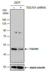
- Experimental details
- Non-transfected (¡V) and transfected (+) 293T whole cell extracts (30 ?g) were separated by 10% SDS-PAGE, and the membrane was blotted with TSG101 antibody (GTX118736) diluted at 1:500. The HRP-conjugated anti-rabbit IgG antibody (GTX213110-01) was used to detect the primary antibody.

Supportive validation
- Submitted by
- GeneTex (provider)
- Main image

- Experimental details
- Sample (30 ug of whole cell lysate) A: A549 10% SDS PAGE GTX118736 diluted at 1:1000
- Validation comment
- WB

- Submitted by
- GeneTex (provider)
- Main image

- Experimental details
- TSG101 antibody detects TSG101 protein by western blot analysis.A. 30 ?g U87-MG whole cell lysate/extractB. 30 ?g SK-N-SH whole cell lysate/extractC. 30 ?g IMR32 whole cell lysate/extractD. 30 ?g SK-N-AS whole cell lysate/extract10 % SDS-PAGETSG101 antibody (GTX118736) dilution: 1:1000
- Validation comment
- WB

- Submitted by
- GeneTex (provider)
- Main image

- Experimental details
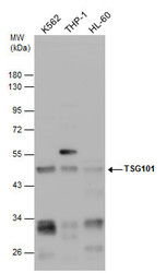
- Various whole cell extracts (30 ?g) were separated by 10% SDS-PAGE, and the membrane was blotted with TSG101 antibody (GTX118736) diluted at 1:500. The HRP-conjugated anti-rabbit IgG antibody (GTX213110-01) was used to detect the primary antibody.

- Submitted by
- GeneTex (provider)
- Main image

- Experimental details
- Various whole cell extracts (30 ?g) were separated by 10% SDS-PAGE, and the membrane was blotted with TSG101 antibody (GTX118736) diluted at 1:1000. The HRP-conjugated anti-rabbit IgG antibody (GTX213110-01) was used to detect the primary antibody.

- Submitted by
- GeneTex (provider)
- Main image
- Experimental details
- Various whole cell extracts (30 ?g) were separated by 10% SDS-PAGE, and the membrane was blotted with TSG101 antibody (GTX118736) diluted at 1:500. The HRP-conjugated anti-rabbite IgG antibody (GTX213110-01) was used to detect the primary antibody, and the signal was developed with Trident femto Western HRP Substrate (GTX14698).

- Submitted by
- GeneTex (provider)
- Main image

- Experimental details
- TSG101 antibody detects TSG101 protein by Western blot analysis.A. 30 £gg U87-MG whole cell lysate/extractB. 30 £gg SK-N-SH whole cell lysate/extractC. 30 £gg IMR32 whole cell lysate/extractD. 30 £gg SK-N-AS whole cell lysate/extract10 % SDS-PAGETSG101 antibody (GTX118736) dilution: 1:500

- Submitted by
- GeneTex (provider)
- Main image

- Experimental details
- TSG101 antibody detects TSG101 protein by Western blot analysis.A. 30 £gg A549 whole cell lysate/extractB. 30 £gg H1299 whole cell lysate/extractC. 30 £gg HCT116 whole cell lysate/extract10 % SDS-PAGETSG101 antibody (GTX118736) dilution: 1:500

- Submitted by
- GeneTex (provider)
- Main image

- Experimental details
- Non-transfected (¡V) and transfected (+) 293T whole cell extracts (30 ?g) were separated by 10% SDS-PAGE, and the membrane was blotted with TSG101 antibody (GTX118736) diluted at 1:500. The HRP-conjugated anti-rabbit IgG antibody (GTX213110-01) was used to detect the primary antibody.

Supportive validation
- Submitted by
- GeneTex (provider)
- Main image
- Experimental details
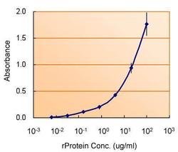
- ELISA detection of TSG101 using GTX118736 for capture at a concentration of 5 ?g/mL and GTX70255 for detection at a concentration of 1.5 ?g/mL.

Supportive validation
- Submitted by
- GeneTex (provider)
- Main image

- Experimental details
- TSG101 antibody detects TSG101 protein at cytoplasm by immunofluorescent analysis. Sample: A549 cells were fixed in 2% paraformaldehyde at RT for 15 min, and be permeabilized in 0.1% saponin, 10% NGS, 100mM glycine for 30 min. Green: TSG101 protein stained by TSG101 antibody (GTX118736) diluted at 1:500. Blue: Hoechst 33342 staining.

Supportive validation
- Submitted by
- GeneTex (provider)
- Main image

- Experimental details
- Immunoprecipitation of TSG101 protein from A549 whole cell extracts using 5 £gg of TSG101 antibody (GTX118736).Western blot analysis was performed using TSG101 antibody (GTX118736).EasyBlot anti-Rabbit IgG (GTX221666-01) was used as a secondary reagent.
