Explore
 Validate
 Learn
Learn Western blot
 Immunocytochemistry
Antibody data
- Antibody Data
- Antigen structure
- References [1]
- Comments [0]
- Validations
- Immunocytochemistry [2]
- Immunohistochemistry [3]
- Flow cytometry [2]
- Other assay [1]
Submit
Validation data
Reference
Comment
Report error
- Product number
- MA5-32687 - Provider product page

- Provider
- Invitrogen Antibodies
- Product name
- MRP2 Recombinant Rabbit Monoclonal Antibody (JA32-01)
- Antibody type
- Monoclonal
- Antigen
- Synthetic peptide
- Reactivity
- Human
- Host
- Rabbit
- Isotype
- IgG
- Antibody clone number
- JA32-01
- Vial size
- 100 µL
- Concentration
- 1 mg/mL
- Storage
- Store at 4°C short term. For long term storage, store at -20°C, avoiding freeze/thaw cycles.
Submitted references Generation of Human Pluripotent Stem Cell-Derived Polarized Hepatocytes.
Bushweller L, Zhao Y, Zhang F, Wu X
Current protocols 2022 Jan;2(1):e345
Current protocols 2022 Jan;2(1):e345
No comments: Submit comment
Supportive validation
- Submitted by
- Invitrogen Antibodies (provider)
- Main image
- Experimental details
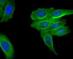
- Immunocytochemical analysis of MRP2 in HepG2 cells using a MRP2 Monoclonal antibody (Product # MA5-32687) as seen in green. The nuclear counter stain is DAPI (blue). Cells were fixed in paraformaldehyde, permeabilised with 0.25% Triton X100/PBS.

- Submitted by
- Invitrogen Antibodies (provider)
- Main image

- Experimental details
- Immunocytochemical analysis of MRP2 in 293T cells using a MRP2 Monoclonal antibody (Product # MA5-32687) as seen in green. The nuclear counter stain is DAPI (blue). Cells were fixed in paraformaldehyde, permeabilised with 0.25% Triton X100/PBS.

Supportive validation
- Submitted by
- Invitrogen Antibodies (provider)
- Main image

- Experimental details
- Immunohistochemical analysis of MRP2 of paraffin-embedded Human liver tissue using a MRP2 Monoclonal antibody (Product #MA5-32687). Counter stained with hematoxylin.

- Submitted by
- Invitrogen Antibodies (provider)
- Main image

- Experimental details
- Immunohistochemical analysis of MRP2 of paraffin-embedded Human kidney tissue using a MRP2 Monoclonal antibody (Product #MA5-32687). Counter stained with hematoxylin.

- Submitted by
- Invitrogen Antibodies (provider)
- Main image

- Experimental details
- Immunohistochemical analysis of MRP2 of paraffin-embedded Human pancreas tissue using a MRP2 Monoclonal antibody (Product #MA5-32687). Counter stained with hematoxylin.

Supportive validation
- Submitted by
- Invitrogen Antibodies (provider)
- Main image

- Experimental details
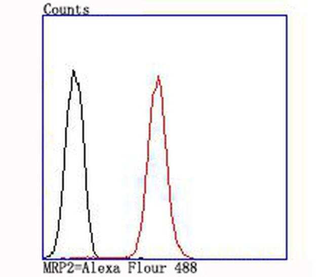
- Flow Cytometric analysis of MRP2 in A549 cells using a MRP2 Monoclonal Antibody (Product # MA5-32687) at a dilution of 1:100, as seen in red compared with an unlabelled control (cells without incubation with primary antibody; black).
- Submitted by
- Invitrogen Antibodies (provider)
- Main image

- Experimental details
- Flow Cytometric analysis of MRP2 in A549 cells using a MRP2 Monoclonal Antibody (Product # MA5-32687) at a dilution of 1:100, as seen in red compared with an unlabelled control (cells without incubation with primary antibody; black).

Supportive validation
- Submitted by
- Invitrogen Antibodies (provider)
- Main image
- Experimental details
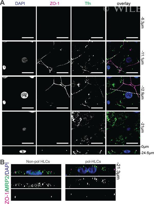
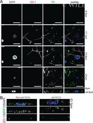
- 4 Figure Distribution of membrane proteins TFRC and MRP2 on pol-HLCs. ( A ) pol-HLCs were incubated with 25 ug/ml of transferrin-488 (green) for 10 min at 37degC prior to washing and staining with anti-ZO-1 (red). Scale bars = 15 mum. Bottom panels: xz images from cross sections indicated by the dashed line in the corresponding xy images above. ( B ) Cross-sectional views ( xz ) of nonpolarized HLCs (non-pol HLCs) and pol-HLCs stained for MRP2 and ZO-1. This figure is adapted from Dao Thi et al. ( 2020).