 Explore
 Validate
 Learn
Learn Western blot
 Immunocytochemistry
Antibody data
- Antibody Data
- Antigen structure
- References [2]
- Comments [0]
- Validations
- Western blot [1]
- Immunohistochemistry [1]
Submit
Validation data
Reference
Comment
Report error
- Product number
- NB100-56087 - Provider product page

- Provider
- Novus Biologicals
- Proper citation
- Novus Cat#NB100-56087, RRID:AB_837675
- Product name
- Rabbit Polyclonal BAG2 Antibody
- Antibody type
- Polyclonal
- Description
- Unpurified. BAG-2
- Reactivity
- Human
- Host
- Rabbit
- Isotype
- IgG
- Vial size
- 0.05 ml
- Storage
- Store at 4C short term. Aliquot and store at -20C long term. Avoid freeze-thaw cycles.
Submitted references Proteome-wide identification of novel binding partners to the oncogenic fusion gene protein, NPM-ALK, using tandem affinity purification and mass spectrometry.
Regulation of the cytoplasmic quality control protein degradation pathway by BAG2.
Wu F, Wang P, Young LC, Lai R, Li L
The American journal of pathology 2009 Feb;174(2):361-70
The American journal of pathology 2009 Feb;174(2):361-70
Regulation of the cytoplasmic quality control protein degradation pathway by BAG2.
Dai Q, Qian SB, Li HH, McDonough H, Borchers C, Huang D, Takayama S, Younger JM, Ren HY, Cyr DM, Patterson C
The Journal of biological chemistry 2005 Nov 18;280(46):38673-81
The Journal of biological chemistry 2005 Nov 18;280(46):38673-81
No comments: Submit comment
Supportive validation
- Submitted by
- Novus Biologicals (provider)
- Main image

- Experimental details
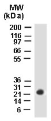
- Western Blot: BAG2 Antibody [NB100-56087] - analysis of BAG-2 using this antibody. HeLa stably transfected Myc-tagged CHIP (
Supportive validation
- Submitted by
- Novus Biologicals (provider)
- Main image
- Experimental details
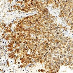
- Immunohistochemistry-Paraffin: BAG2 Antibody [NB100-56087] - BAG-2 was detected in immersion fixed paraffin-embedded sections of human testis using Rabbit Anti-Human BAG-2 polyclonal Antibody (Catalog # NB100-56087) at 1:1000 for 1 hour at room temperature followed by incubation with the Anti-Rabbit IgG VisUCyte™ HRP Polymer Antibody (Catalog # VC003). Tissue was stained using DAB (brown) and counterstained with hematoxylin (blue). Specific staining was localized to the cytoplasm and plasma membrane.
