 Explore
 Validate
 Learn
Learn Western blot
 Immunohistochemistry
Antibody data
- Antibody Data
- Antigen structure
- References [1]
- Comments [0]
- Validations
- Western blot [1]
- Immunocytochemistry [2]
- Immunohistochemistry [14]
Submit
Validation data
Reference
Comment
Report error
- Product number
- HPA006385 - Provider product page

- Provider
- Atlas Antibodies
- Proper citation
- Atlas Antibodies Cat#HPA006385, RRID:AB_1080447
- Product name
- Anti-UBTF
- Antibody type
- Polyclonal
- Reactivity
- Human, Mouse
- Host
- Rabbit
- Conjugate
- Unconjugated
- Antigen sequence
WKLLSQKEKDAYHKKCDQKKKDYEVELLRFLESLP
EEEQQRVLGEEKMLNINKKQATSPASKKPAQEGGK
GGSEKPKRPVSAMFIFSEEKRRQLQEERPELSESE
LTRLLARMWNDLSEKKK- Isotype
- IgG
- Vial size
- 100 µl
- Storage
- Store at +4°C for short term storage. Long time storage is recommended at -20°C.
Submitted references UBF complexes with phosphatidylinositol 4,5-bisphosphate in nucleolar organizer regions regardless of ongoing RNA polymerase I activity.
Sobol M, Yildirim S, Philimonenko VV, Marášek P, Castaño E, Hozák P
Nucleus (Austin, Tex.) 2013 Nov-Dec;4(6):478-86
Nucleus (Austin, Tex.) 2013 Nov-Dec;4(6):478-86
No comments: Submit comment
Enhanced validation
- Submitted by
- Atlas Antibodies (provider)
- Enhanced method
- Genetic validation
- Main image

- Experimental details
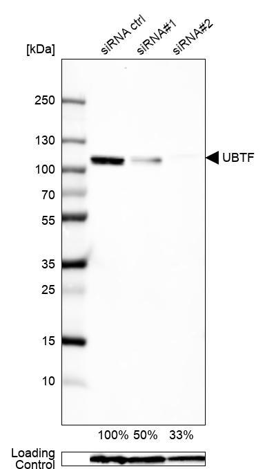
- Western blot analysis in U2OS cells transfected with control siRNA, target specific siRNA probe #1 and #2, using Anti-UBTF antibody. Remaining relative intensity is presented. Loading control: Anti-GAPDH.
Enhanced validation
Supportive validation
- Submitted by
- 55af80e3e0991
- Enhanced method
- Genetic validation
- Main image
- Experimental details
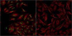
- Confocal images of immunofluorescently stained human U-2 OS cells.The protein UBTF is shown in green and the microtubules in red. The image to the left show cells transfected with control siRNA and the image to the right show cells where UBTF has been downregulated with specific siRNA.
- Sample type
- U-2 OS cells
- Primary Ab dilution
- 1:72
- Secondary Ab
- Secondary Ab
- Secondary Ab dilution
- 1:800
- Knockdown/Genetic Approaches Application
- Immunocytochemistry

Supportive validation
- Submitted by
- Atlas Antibodies (provider)
- Main image
- Experimental details
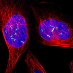
- Immunofluorescent staining of human cell line U-2 OS shows localization to nucleoli fibrillar center.
- Sample type
- HUMAN

Supportive validation
- Submitted by
- Atlas Antibodies (provider)
- Main image
- Experimental details
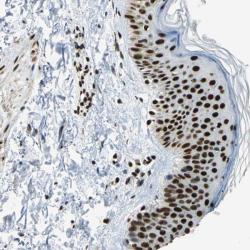
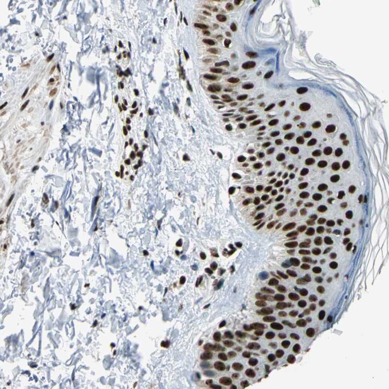
- Immunohistochemical staining of human skin shows strong nuclear positivity in epidermal cells.
- Sample type
- HUMAN
- Submitted by
- Atlas Antibodies (provider)
- Main image
- Experimental details
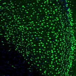
- Immunofluorescence staining of mouse retrosplenial granular cortex shows nuclear neuronal positivity.
- Sample type
- MOUSE

- Submitted by
- Atlas Antibodies (provider)
- Main image
- Experimental details
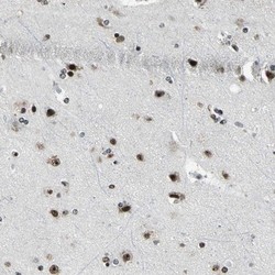
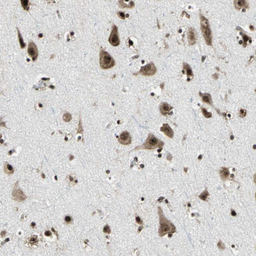
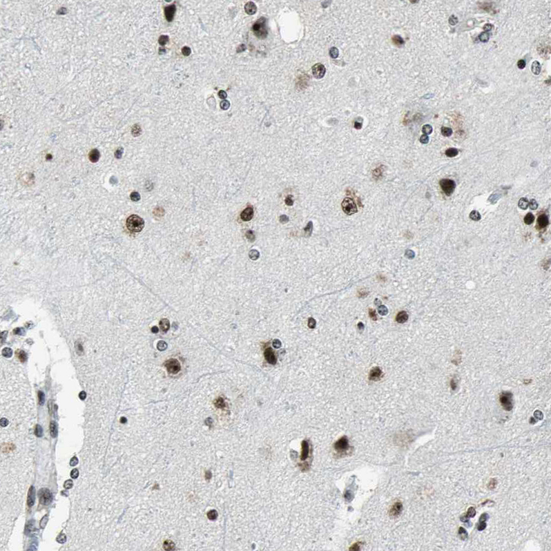
- Immunohistochemical staining of human cerebral cortex shows nuclear positivity.
- Sample type
- HUMAN

- Submitted by
- Atlas Antibodies (provider)
- Main image

- Experimental details
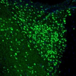
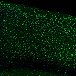
- Immunofluorescence staining of mouse motor cortex shows nuclear positivity in neurons.
- Sample type
- MOUSE

- Submitted by
- Atlas Antibodies (provider)
- Main image
- Experimental details
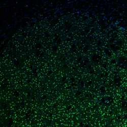
- Immunofluorescence staining of mouse thalamus shows nuclear immunoreactivity in anterodorsal thalamic nucleus neurons.
- Sample type
- MOUSE

- Submitted by
- Atlas Antibodies (provider)
- Main image

- Experimental details
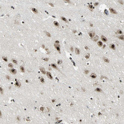
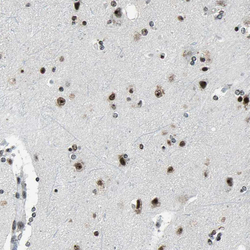
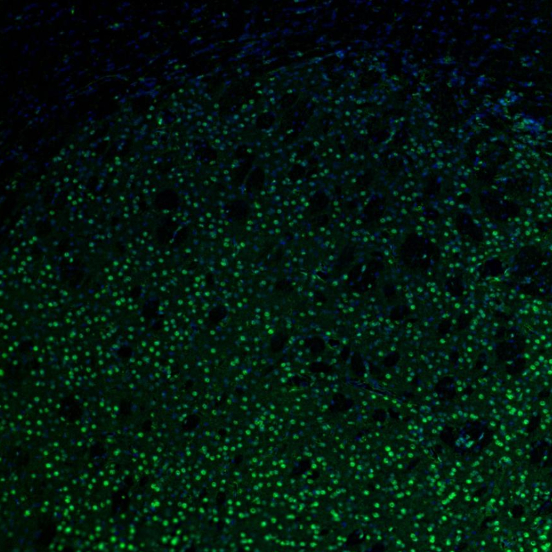
- Immunohistochemical staining of human cerebral cortex shows nuclear and cytoplasmic immunoreactivity in neurons.
- Submitted by
- Atlas Antibodies (provider)
- Main image
- Experimental details
- Immunohistochemical staining of human hippocampus shows nuclear immunoreactivity in neurons.
- Sample type
- HUMAN

- Submitted by
- Atlas Antibodies (provider)
- Main image
- Experimental details
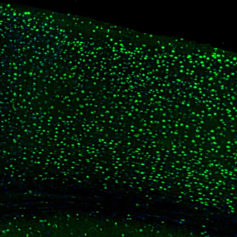
- Immunofluorescence staining of mouse brain shows strong nuclear positivity in neurons in the cerebral cortex.
- Sample type
- MOUSE
- Submitted by
- Atlas Antibodies (provider)
- Main image

- Experimental details
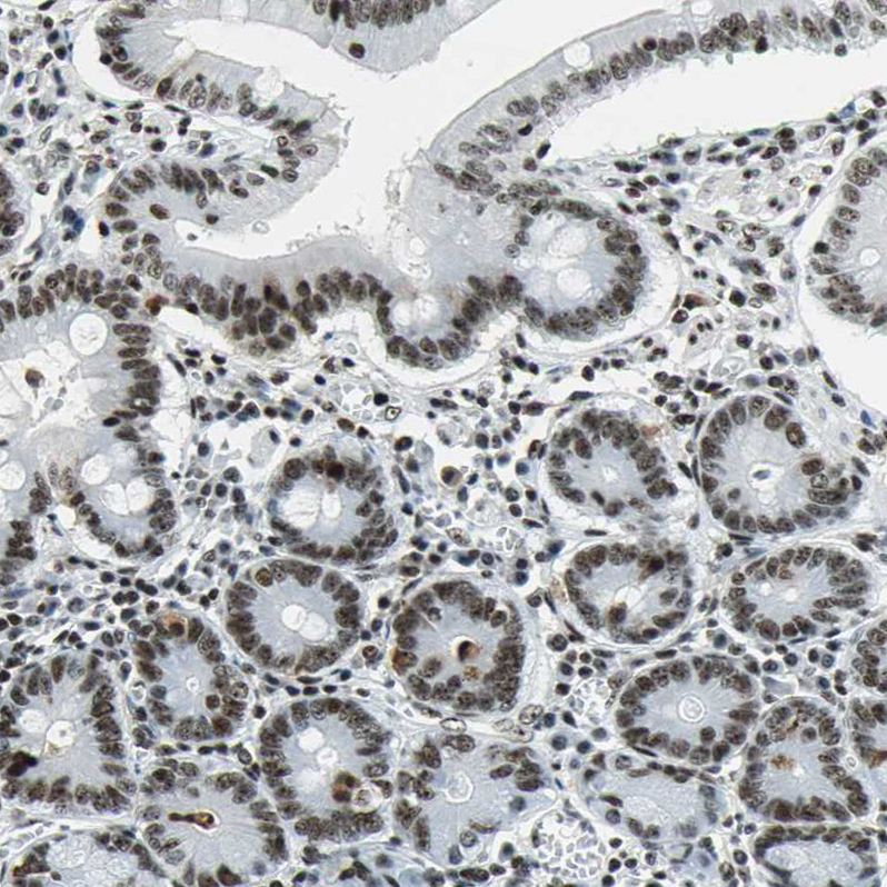
- Immunohistochemical staining of human small intestine shows moderate nuclear positivity in glandular cells.
- Sample type
- HUMAN
- Submitted by
- Atlas Antibodies (provider)
- Main image
- Experimental details
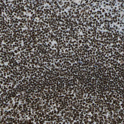
- Immunohistochemical staining of human tonsil shows strong nuclear positivity in non-germinal center cells.
- Sample type
- HUMAN

- Submitted by
- Atlas Antibodies (provider)
- Main image
- Experimental details
- Immunohistochemical staining of human cerebral cortex shows moderate to strong nuclear positivity in neurons.
- Sample type
- HUMAN
- Submitted by
- Atlas Antibodies (provider)
- Main image
- Experimental details
- Immunofluorescence staining of mouse basal forebrain shows moderate nuclear positivity in neurons in the caudate putamen.
- Sample type
- MOUSE
- Submitted by
- Atlas Antibodies (provider)
- Main image
- Experimental details
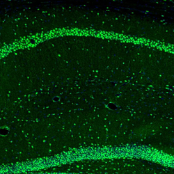
- Immunofluorescence staining of mouse brain shows strong positivity in neurons in the CA1 and granular cell layers in the hippocampus.
- Sample type
- MOUSE

- Submitted by
- Atlas Antibodies (provider)
- Main image
- Experimental details
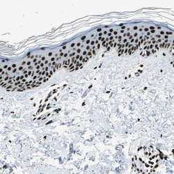
- Immunohistochemical staining of human skin shows strong nuclear positivity in squamous epithelial cells.
- Sample type
- HUMAN
