 Explore
 Validate
 Learn
Learn Western blot
 Other assay
Antibody data
- Antibody Data
- Antigen structure
- References [3]
- Comments [0]
- Validations
- Other assay [2]
Submit
Validation data
Reference
Comment
Report error
- Product number
- PA1-941 - Provider product page

- Provider
- Invitrogen Antibodies
- Product name
- KCNQ5 Polyclonal Antibody
- Antibody type
- Polyclonal
- Antigen
- Synthetic peptide
- Description
- PA1-941 detects the KCNQ5 from rat and human samples. This antibody is specific for KCNQ5 and does not detect KCNQ1, KCNQ2, KCNQ3 or KCNQ4. PA1-941 has been successfully used in Western blot procedures. By Western blot, this antibody detects a ~125 kDa band representing KCNQ5 from rat brain extract. The PA1-941 immunizing peptide corresponds to amino acid residues 880-897 from human KCNQ5.
- Reactivity
- Human, Rat
- Host
- Rabbit
- Isotype
- IgG
- Vial size
- 200 µg
- Concentration
- 1 mg/mL
- Storage
- -20° C, Avoid Freeze/Thaw Cycles
Submitted references Peripheral K(V)7 channels regulate visceral sensory function in mouse and human colon.
Distribution of voltage-gated potassium and hyperpolarization-activated channels in sensory afferent fibers in the rat carotid body.
The KCNQ/M-current modulates arterial baroreceptor function at the sensory terminal in rats.
Peiris M, Hockley JR, Reed DE, Smith ESJ, Bulmer DC, Blackshaw LA
Molecular pain 2017 Jan-Dec;13:1744806917709371
Molecular pain 2017 Jan-Dec;13:1744806917709371
Distribution of voltage-gated potassium and hyperpolarization-activated channels in sensory afferent fibers in the rat carotid body.
Buniel M, Glazebrook PA, Ramirez-Navarro A, Kunze DL
The Journal of comparative neurology 2008 Oct 1;510(4):367-77
The Journal of comparative neurology 2008 Oct 1;510(4):367-77
The KCNQ/M-current modulates arterial baroreceptor function at the sensory terminal in rats.
Wladyka CL, Feng B, Glazebrook PA, Schild JH, Kunze DL
The Journal of physiology 2008 Feb 1;586(3):795-802
The Journal of physiology 2008 Feb 1;586(3):795-802
No comments: Submit comment
Supportive validation
- Submitted by
- Invitrogen Antibodies (provider)
- Main image
- Experimental details
- NULL

- Submitted by
- Invitrogen Antibodies (provider)
- Main image

- Experimental details
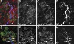
- Figure 1. Expression of K V 7 channels in mouse colon and mesenteric neurovasculature. (a) K V 7.3 co-expression with CGRP-positive neurons within the myenteric plexus (MP) of mouse colon (tissue sections). Panel (iv) is a schematic representation of endings that co-label for CGRP and K V 7.3 (in black) surrounding neuronal cell bodies (in grey) of the myenteric plexus. (b) In the mesentery associated with the colon, K V 7.3 co-localizes with CGRP-positive neurons (white arrowheads) surrounding blood vessels (BV) in whole mounts. (c) K V 7.5 is expressed in the serosal layer of mouse colon and co-localizes with CGRP-positive fibres in the myenteric plexus (tissue sections). (d) CGRP positive neurons that run alongside blood vessels found within the mesenteric attachment to the colon co-express K V 7.5 (whole mounts). Punctate staining for K V 7.5 is also found around CGRP positive nerve bundle (NF). All images taken at 40X, scale bar = 25 um. Figures b(iii) and d(iii) are 4X of the original images, b(i) and d(i). LM: longitudinal muscle; CM: circular muscle.
