 Explore
 Validate
 Learn
Learn Western blot
 Immunohistochemistry
Antibody data
- Antibody Data
- Antigen structure
- References [6]
- Comments [0]
- Validations
- Immunohistochemistry [1]
- Other assay [2]
Submit
Validation data
Reference
Comment
Report error
- Product number
- 417200 - Provider product page

- Provider
- Invitrogen Antibodies
- Product name
- FXR Monoclonal Antibody (A9033A)
- Antibody type
- Monoclonal
- Antigen
- Recombinant full-length protein
- Reactivity
- Human, Mouse, Rat
- Host
- Mouse
- Isotype
- IgG
- Antibody clone number
- A9033A
- Vial size
- 100 µL
- Concentration
- 1 mg/mL
- Storage
- Maintain refrigerated at 2-8°C for up to 1 month. For long term storage store at -20°C
Submitted references Pregnancy and weaning regulate human maternal liver size and function.
Hyperoside attenuates non-alcoholic fatty liver disease in rats via cholesterol metabolism and bile acid metabolism.
Gypenosides regulate farnesoid X receptor-mediated bile acid and lipid metabolism in a mouse model of non-alcoholic steatohepatitis.
The Status of Bile Acids and Farnesoid X Receptor in Brain and Liver of Rats with Thioacetamide-Induced Acute Liver Failure.
FXR Isoforms Control Different Metabolic Functions in Liver Cells via Binding to Specific DNA Motifs.
Bile acids induce hepatic differentiation of mesenchymal stem cells.
Q Bartlett A, Vesco KK, Purnell JQ, Francisco M, Goddard E, Guan X, DeBarber A, Leo MC, Baetscher E, Rooney W, Naugler W, Guimaraes AR, Catalano P, Xia Z, Schedin P
Proceedings of the National Academy of Sciences of the United States of America 2021 Nov 30;118(48)
Proceedings of the National Academy of Sciences of the United States of America 2021 Nov 30;118(48)
Hyperoside attenuates non-alcoholic fatty liver disease in rats via cholesterol metabolism and bile acid metabolism.
Wang S, Sheng F, Zou L, Xiao J, Li P
Journal of advanced research 2021 Dec;34:109-122
Journal of advanced research 2021 Dec;34:109-122
Gypenosides regulate farnesoid X receptor-mediated bile acid and lipid metabolism in a mouse model of non-alcoholic steatohepatitis.
Li H, Xi Y, Xin X, Tian H, Hu Y
Nutrition & metabolism 2020;17:34
Nutrition & metabolism 2020;17:34
The Status of Bile Acids and Farnesoid X Receptor in Brain and Liver of Rats with Thioacetamide-Induced Acute Liver Failure.
Czarnecka AM, Milewski K, Albrecht J, Zielińska M
International journal of molecular sciences 2020 Oct 20;21(20)
International journal of molecular sciences 2020 Oct 20;21(20)
FXR Isoforms Control Different Metabolic Functions in Liver Cells via Binding to Specific DNA Motifs.
Ramos Pittol JM, Milona A, Morris I, Willemsen ECL, van der Veen SW, Kalkhoven E, van Mil SWC
Gastroenterology 2020 Nov;159(5):1853-1865.e10
Gastroenterology 2020 Nov;159(5):1853-1865.e10
Bile acids induce hepatic differentiation of mesenchymal stem cells.
Sawitza I, Kordes C, Götze S, Herebian D, Häussinger D
Scientific reports 2015 Aug 25;5:13320
Scientific reports 2015 Aug 25;5:13320
No comments: Submit comment
Supportive validation
- Submitted by
- Invitrogen Antibodies (provider)
- Main image
- Experimental details
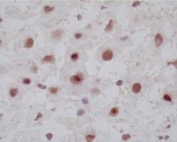
- Immunohistochemistry analysis of FRX in frozen rat liver tissues. The sample was inclubated in FRX monoclonal antibody (Product # 417200) at a dilution of 3~20 μg/mL. Section was fixed with 4% paraformaldehyde-fixed in immersion, activated with 5% TritonX-100 for 5 minutes, and blocked with 3% H2O2 in methanol.
Supportive validation
- Submitted by
- Invitrogen Antibodies (provider)
- Main image
- Experimental details
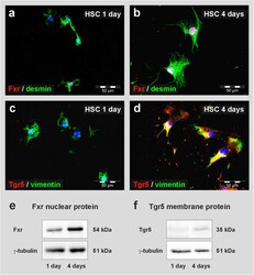
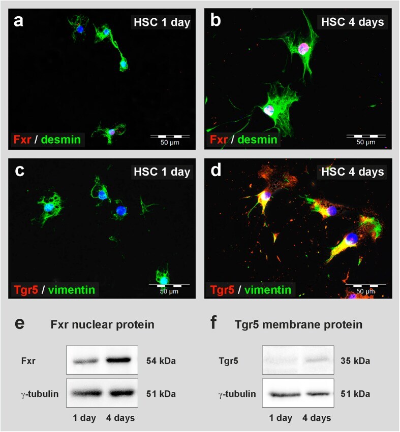
- Figure 6 Detection of bile acid receptors in freshly isolated and activated HSC from rats. Freshly isolated (1 day) and cultured (4 days) HSC were stained with antibodies against the bile acid receptors ( a,b ) Fxr and ( c,d ) Tgr5 (red). The mesenchymal markers desmin and vimentin (green) were used to verify the presence of stellate cells. HSC cultured for 4 days were maintained in medium without serum and without TUDCA. ( a ) Few freshly isolated HSC displayed weak nuclear staining of Fxr, but most cells lacked clear nuclear Fxr staining one day after isolation. ( b ) The majority of HSC exhibited significant nuclear Fxr after few days in culture. ( c ) Tgr5 was not detectable in freshly isolated HSC by immunofluorescence, ( d ) but appeared during culture within few days. ( e ) The presence of Fxr in freshly isolated and cultured HSC was confirmed by Western blot analysis. Fxr was detected in the nuclear protein faction mainly after 4 days of culture (10 mug protein per lane). ( f ) Tgr5 was found in cell membrane protein fraction of HSC after 4 days of culture (30 mug protein per lane). The protein gamma-tubulin served as a control.
- Submitted by
- Invitrogen Antibodies (provider)
- Main image
- Experimental details
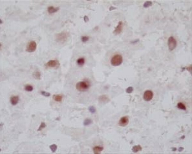
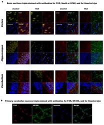
- Figure 1 ( a ) Immunolabelling of farnesoid X receptor, FXR (red) in rat cerebral cortex, hippocampus, and cerebellum of control and thioacetamide (TAA) rats. Neurons were positively immunostained against the neuronal marker NeuN (yellow). Astrocytes were identified by immunostaining against the glial fibrillary acidic protein (GFAP, green). Blue Hoechst stain indicates the location of cell nuclei. Dashed lines indicate the Purkinje layer. Scale bars = 10 mum; ( b ) immunolabelling of FXR in the primary cultures of rat cerebellar neurons. Neurons were positively immunostained against the neuronal marker neurofilament 200 (NF200, green). Blue Hoechst stain indicates the location of cell nuclei. Scale bars = 10 mum.
