 Explore
 Validate
 Learn
Learn Western blot
 Immunocytochemistry
Antibody data
- Antibody Data
- Antigen structure
- References [0]
- Comments [0]
- Validations
- Western blot [4]
- Immunoprecipitation [1]
- Immunohistochemistry [1]
Submit
Validation data
Reference
Comment
Report error
- Product number
- NBP2-19499 - Provider product page

- Provider
- Novus Biologicals
- Product name
- Rabbit Polyclonal NDP52 Antibody
- Antibody type
- Polyclonal
- Description
- Immunogen affinity purified.
- Reactivity
- Human, Mouse
- Host
- Rabbit
- Isotype
- IgG
- Vial size
- 0.1 ml
- Storage
- Aliquot and store at -20C or -80C. Avoid freeze-thaw cycles.
No comments: Submit comment
Supportive validation
- Submitted by
- Novus Biologicals (provider)
- Main image

- Experimental details
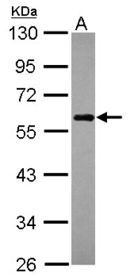
- Western Blot: NDP52 Antibody [NBP2-19499] - Sample (30 ug of whole cell lysate) A: Raji 10% SDS PAGE gel, diluted at 1:1000.
- Submitted by
- Novus Biologicals (provider)
- Main image
- Experimental details
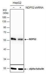
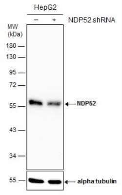
- Western Blot: NDP52 Antibody [NBP2-19499] - Non-transfected (-) and transfected (+) HepG2 whole cell extracts (30 ug) were separated by 10% SDS-PAGE, and the membrane was blotted with NDP52 antibody. HRP-conjugated anti-rabbit IgG antibody was used to detect the primary antibody.
- Submitted by
- Novus Biologicals (provider)
- Main image
- Experimental details
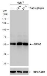
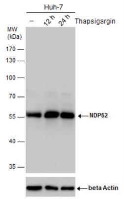
- Western Blot: NDP52 Antibody [NBP2-19499] - NDP52 antibody detects NDP52 protein by western blot analysis. Un-treated (-) and treated (+), Thapsigargin treatment for 12hrs and 24hrs. Huh-7 whole cell extracts (30 ug) were separated by 10% SDS-PAGE, and the membrane was blotted with NDP52 antibody. ACTB was used as internal control, shown at the bottom panel. HRP-conjugated anti-rabbit IgG antibody was used to detect the primary antibody.
- Submitted by
- Novus Biologicals (provider)
- Main image

- Experimental details
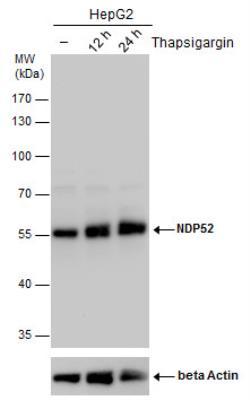
- Western Blot: NDP52 Antibody [NBP2-19499] - NDP52 antibody detects NDP52 protein by western blot analysis. Un-treated (-) and treated (+, Thapsigargin treatment for 12hrs and 24hrs) HepG2 whole cell extracts (30 ug) were separated by 10% SDS-PAGE, and the membrane was blotted with NDP52 antibody. The ACTB was used as internal control, shown at the bottom panel. HRP-conjugated anti-rabbit IgG antibody was used to detect the primary antibody.
Supportive validation
- Submitted by
- Novus Biologicals (provider)
- Main image

- Experimental details
- Immunoprecipitation: NDP52 Antibody [NBP2-19499] - Jurkat whole cell extracts using 5 ug of NDP52 antibody. Western blot analysis was performed using NDP52 antibody. EasyBlot anti-Rabbit IgG was used as a secondary reagent.

Supportive validation
- Submitted by
- Novus Biologicals (provider)
- Main image
- Experimental details
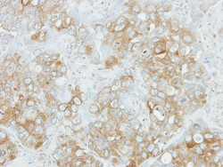
- Immunohistochemistry-Paraffin: NDP52 Antibody [NBP2-19499] - Immunohistochemical analysis of paraffin-embedded H441 xenograft, using antibody at 1:100 dilution.
