 Explore
 Validate
 Learn
Learn Western blot
 Immunoprecipitation
Antibody data
- Antibody Data
- Antigen structure
- References [0]
- Comments [0]
- Validations
- Western blot [4]
- Immunocytochemistry [2]
- Immunohistochemistry [1]
- Other assay [1]
Submit
Validation data
Reference
Comment
Report error
- Product number
- PA5-30367 - Provider product page

- Provider
- Invitrogen Antibodies
- Product name
- CALCOCO2 Polyclonal Antibody
- Antibody type
- Polyclonal
- Antigen
- Recombinant protein fragment
- Description
- Recommended positive controls: Raji, HepG2, HepG2 whole cell extract (untreated), HepG2 whole cell extract (3 µM Thapsigargin treatment for 12 hr), HepG2 whole cell extract (3 µM Thapsigargin treatment for 24 hr), Huh-7 (untreated), Huh-7 (3 µM Thapsigargin treatment for 12 hr), Huh-7 (3 µM Thapsigargin treatment for 24 hr). Predicted reactivity: Human (99%), Chimpanzee (97%). Store product as a concentrated solution. Centrifuge briefly prior to opening the vial.
- Reactivity
- Human, Mouse
- Host
- Rabbit
- Isotype
- IgG
- Vial size
- 100 µL
- Concentration
- 1.57 mg/mL
- Storage
- Store at 4°C short term. For long term storage, store at -20°C, avoiding freeze/thaw cycles.
No comments: Submit comment
Supportive validation
- Submitted by
- Invitrogen Antibodies (provider)
- Main image
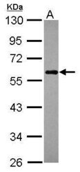
- Experimental details
- Western Blot using CALCOCO2 Polyclonal Antibody (Product # PA5-30367). Sample (30 µg of whole cell lysate). Lane A: Raji. 10% SDS PAGE. CALCOCO2 Polyclonal Antibody (Product # PA5-30367) diluted at 1:1,000. The HRP-conjugated anti-rabbit IgG antibody was used to detect the primary antibody.

- Submitted by
- Invitrogen Antibodies (provider)
- Main image
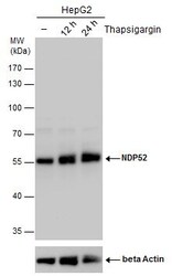
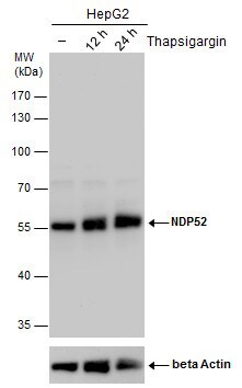
- Experimental details
- CALCOCO2 Polyclonal Antibody detects NDP52 protein by western blot analysis. Un-treated (-) and treated (+, Thapsigargin treatment for 12hrs and 24hrs) HepG2 whole cell extracts (30 µg) were separated by 10% SDS-PAGE, and the membrane was blotted with CALCOCO2 Polyclonal Antibody (Product # PA5-30367) diluted by 1:2,000. The ACTB was used as internal control (Product # MA1-20221)4, 1:50,000) shown at the bottom panel. The HRP-conjugated anti-rabbit IgG antibody was used to detect the primary antibody.
- Submitted by
- Invitrogen Antibodies (provider)
- Main image
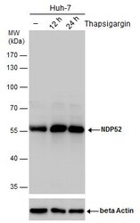
- Experimental details
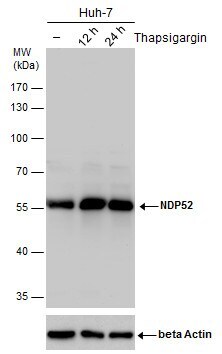
- CALCOCO2 Polyclonal Antibody detects NDP52 protein by western blot analysis. Un-treated (-) and treated (+, Thapsigargin treatment for 12hrs and 24hrs) Huh-7 whole cell extracts (30 µg) were separated by 10% SDS-PAGE, and the membrane was blotted with CALCOCO2 Polyclonal Antibody (Product # PA5-30367) diluted by 1:2,000. The ACTB was used as internal control (Product # MA1-20221)4, 1:50,000) shown at the bottom panel. The HRP-conjugated anti-rabbit IgG antibody was used to detect the primary antibody.
- Submitted by
- Invitrogen Antibodies (provider)
- Main image
- Experimental details
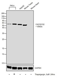
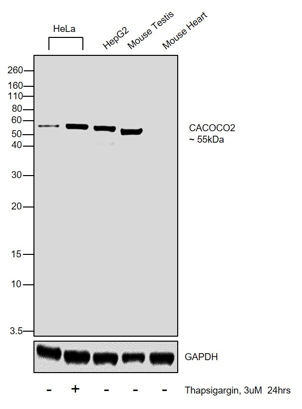
- Western blot was performed using Anti-CALCOCO2 Polyclonal Antibody (Product # PA5-30367) and a 55kDa band corresponding to CALCOCO2 was observed across the cell lines tested and tissues tested. Whole cell extracts (30 µg lysate) of HeLa (Lane 1), HeLa treated with Thapsigargin (3uM for 24hrs) (Lane 2), Hep G2 (Lane 3), Mouse Testis (Lane 4), Mouse Heart (Lane 5) were electrophoresed using NuPAGE™ 12% Bis-Tris Protein Gel (Product # NP0341BOX). Resolved proteins were then transferred onto a Nitrocellulose membrane (Product # IB23001) by iBlot® 2 Dry Blotting System (Product # IB21001). The blot was probed with the primary antibody (1:1000) and detected by chemiluminescence with Goat anti-Rabbit IgG (H+L) Superclonal™ Recombinant Secondary Antibody, HRP (Product # A27036, 1:4000) using the iBright FL 1000 (Product # A32752). Chemiluminescent detection was performed using Novex® ECL Chemiluminescent Substrate Reagent Kit (Product # WP20005).
Supportive validation
- Submitted by
- Invitrogen Antibodies (provider)
- Main image

- Experimental details
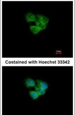
- Immunofluorescent analysis of CALCOCO2 in methanol-fixed A431 cells using a CALCOCO2 polyclonal antibody (Product # PA5-30367) at a 1:200 dilution.
- Submitted by
- Invitrogen Antibodies (provider)
- Main image
- Experimental details
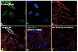
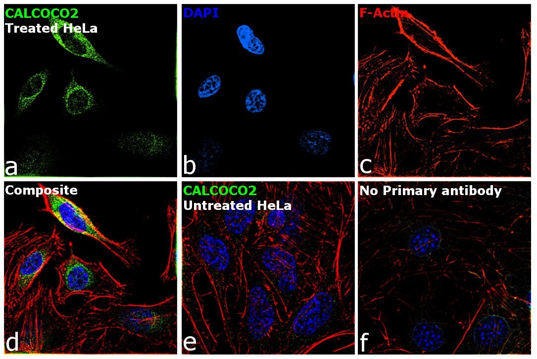
- Immunofluorescence analysis of CALCOCO2 was performed using 70% confluent log phase HeLa cells treated with 3 Thapsigargin 3uM for 48 hrs. The cells were fixed with 4% paraformaldehyde for 10 minutes, permeabilized with 0.1% Triton™ X-100 for 15 minutes, and blocked with 2% BSA for 45 minutes at room temperature. The cells were labeled with CALCOCO2 Polyclonal Antibody (Product # PA5-30367) at 1:100 in 0.1% BSA, incubated at 4 degree celsius overnight and then labeled with Goat anti-Rabbit IgG (H+L) Highly Cross-Adsorbed Secondary Antibody, Alexa Fluor Plus 488 (Product # A32731), (1:2000), for 45 minutes at room temperature (Panel a: Green). Nuclei (Panel b: Blue) were stained with ProLong™ Diamond Antifade Mountant with DAPI (Product # P36962). F-actin (Panel c: Red) was stained with Rhodamine Phalloidin (Product # R415, 1:300). Panel d represents the merged image showing Cytosol localization. Panel e represents untreated HeLa cells showing no signal Panel f represents control cells with no primary antibody to assess background. The images were captured at 60X magnification.
Supportive validation
- Submitted by
- Invitrogen Antibodies (provider)
- Main image
- Experimental details
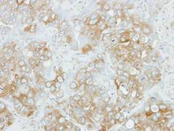
- Immunohistochemical analysis of paraffin-embedded H441 xenograft, using NDP52 (Product # PA5-30367) antibody at 1:100 dilution. Antigen Retrieval: EDTA based buffer, pH 8.0, 15 min.

Supportive validation
- Submitted by
- Invitrogen Antibodies (provider)
- Main image
- Experimental details
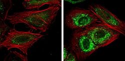
- Immunofluorescent analysis of CALCOCO2 showing staining in the autophagosome of HeLa cells. HeLa cells mock (left) and treated with 50µM Chloroquine for 24 hr (right) were fixed in 4% paraformaldehyde at RT for 15 min and stained using a CALCOCO2 polyclonal antibody (Product # PA5-30367) diluted at 1:1000. Red: Phalloidin, a F-actin marker.
