 Explore
 Validate
 Learn
Learn Western blot
Antibody data
- Antibody Data
- Antigen structure
- References [1]
- Comments [0]
- Validations
- Western blot [3]
- Immunocytochemistry [2]
- Immunohistochemistry [12]
Submit
Validation data
Reference
Comment
Report error
- Product number
- HPA021232 - Provider product page

- Provider
- Atlas Antibodies
- Proper citation
- Atlas Antibodies Cat#HPA021232, RRID:AB_1845837
- Product name
- Anti-C9orf78
- Antibody type
- Polyclonal
- Reactivity
- Human
- Host
- Rabbit
- Conjugate
- Unconjugated
- Antigen sequence
YHEELNAPIRRNKEEPKARPLRVGDTEKPEPERSP
PNRKRPANEKATDDYHYEKFK- Isotype
- IgG
- Vial size
- 100 µl
- Storage
- Store at +4°C for short term storage. Long time storage is recommended at -20°C.
Submitted references Contribution of Antibody-based Protein Profiling to the Human Chromosome-centric Proteome Project (C-HPP)
Fagerberg L, Oksvold P, Skogs M, Älgenäs C, Lundberg E, Pontén F, Sivertsson Å, Odeberg J, Klevebring D, Kampf C, Asplund A, Sjöstedt E, Al-Khalili Szigyarto C, Edqvist P, Olsson I, Rydberg U, Hudson P, Ottosson Takanen J, Berling H, Björling L, Tegel H, Rockberg J, Nilsson P, Navani S, Jirström K, Mulder J, Schwenk J, Zwahlen M, Hober S, Forsberg M, von Feilitzen K, Uhlén M
Journal of Proteome Research 2013 June;12(6):2439-2448
Journal of Proteome Research 2013 June;12(6):2439-2448
No comments: Submit comment
Enhanced validation
Supportive validation
- Submitted by
- klas2
- Enhanced method
- Genetic validation
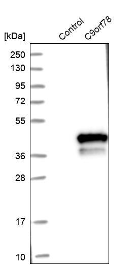
- Main image

- Experimental details
- Western blot of cell lysate from U-2 OS cells transfected with either siRNA targeting C9orf78 or control siRNA. Lane 1: Marker (250, 130, 95, 72, 55, 36, 28, 17, 10) Lane 2: Cell lysate from U-2OS cells transfected with siRNA targeting C9orf78 Lane 3: N/A Lane 4: Cell lysate from U-2OS cells transfected with control siRNA Right image, lane 1-4: loading control
- Sample type
- U-2 OS
- Primary Ab dilution
- 1:58
- Conjugate
- Horseradish Peroxidase
- Secondary Ab
- Secondary Ab
- Secondary Ab dilution
- 1:3000
- Knockdown/Genetic Approaches Application
- Western blot

Supportive validation
- Submitted by
- Atlas Antibodies (provider)
- Enhanced method
- Genetic validation
- Main image
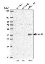
- Experimental details
- Western blot analysis in U2OS cells transfected with control siRNA, target specific siRNA probe #1 and #2, using Anti-C9orf78 antibody. Remaining relative intensity is presented.

- Submitted by
- Atlas Antibodies (provider)
- Enhanced method
- Recombinant expression validation
- Main image
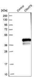
- Experimental details
- Western blot analysis in control (vector only transfected HEK293T lysate) and C9orf78 over-expression lysate (Co-expressed with a C-terminal myc-DDK tag (~3.1 kDa) in mammalian HEK293T cells, LY413954).
Enhanced validation
Supportive validation
- Submitted by
- 55af80e3e0991
- Enhanced method
- Genetic validation
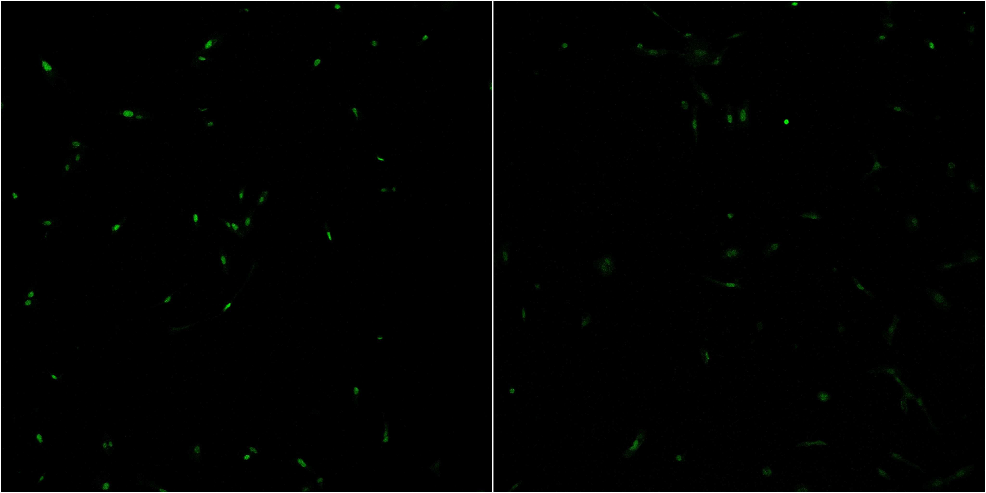
- Main image

- Experimental details
- Confocal images of immunofluorescently stained human U-2 OS cells.The protein C9orf78 is shown in green. The image to the left show cells transfected with control siRNA and the image to the right show cells where C9orf78 has been downregulated with specific siRNA.
- Sample type
- U-2 OS cells
- Primary Ab dilution
- 1:7
- Secondary Ab
- Secondary Ab
- Secondary Ab dilution
- 1:800
- Knockdown/Genetic Approaches Application
- Immunocytochemistry
Supportive validation
- Submitted by
- Atlas Antibodies (provider)
- Main image

- Experimental details
- Immunofluorescent staining of human cell line U-2 OS shows localization to nucleoplasm.
- Sample type
- HUMAN

Enhanced validation
Enhanced validation
Supportive validation
- Submitted by
- Atlas Antibodies (provider)
- Enhanced method
- Orthogonal validation
- Main image

- Experimental details
- Immunohistochemistry analysis in human bone marrow and pancreas tissues using HPA021232 antibody. Corresponding C9orf78 RNA-seq data are presented for the same tissues.
- Sample type
- HUMAN

Enhanced validation
- Submitted by
- Atlas Antibodies (provider)
- Enhanced method
- Independent antibody validation
- Main image

- Experimental details
- Immunohistochemical staining of human bone marrow, fallopian tube, lymph node and testis using Anti-C9orf78 antibody HPA021232 (A) shows similar protein distribution across tissues to independent antibody HPA021216 (B).

Supportive validation
- Submitted by
- Atlas Antibodies (provider)
- Main image

- Experimental details
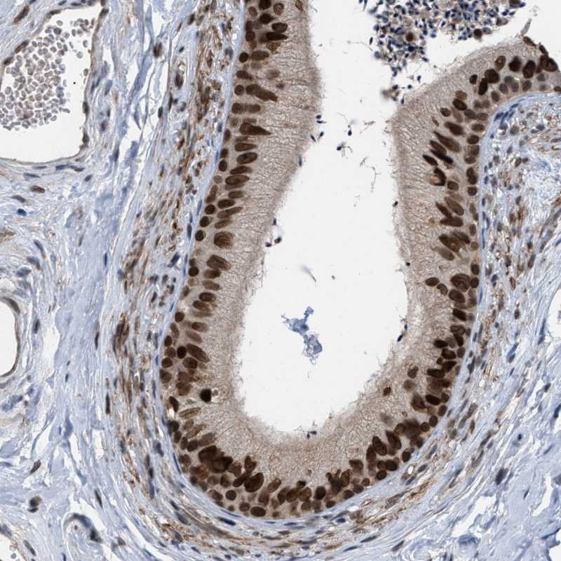
- Immunohistochemical staining of human epididymis shows strong nuclear positivity in glandular cells.
- Submitted by
- Atlas Antibodies (provider)
- Main image

- Experimental details
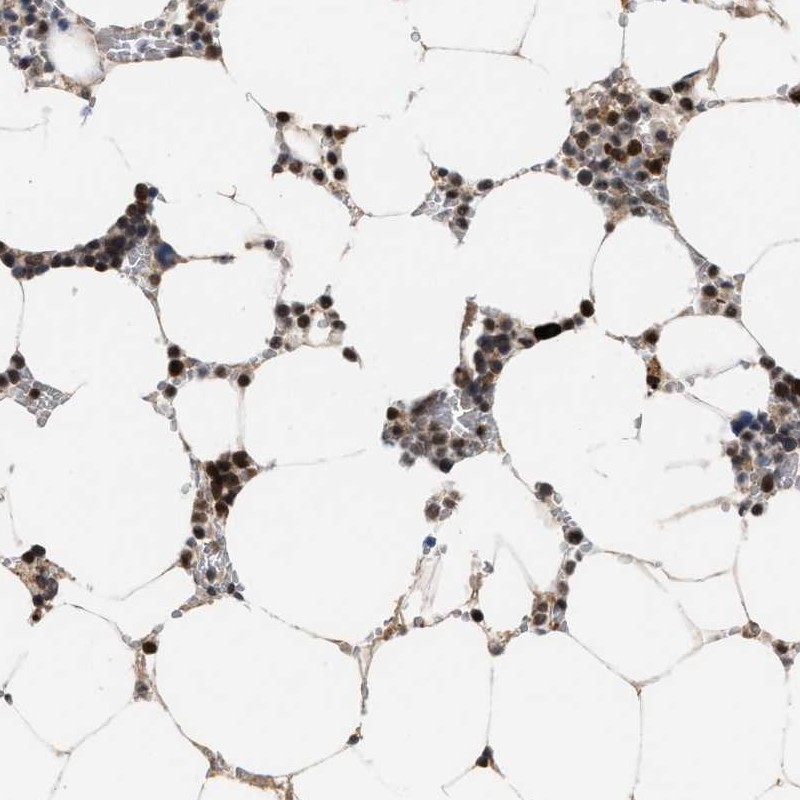
- Immunohistochemical staining of human bone marrow shows high expression.
- Sample type
- HUMAN
- Submitted by
- Atlas Antibodies (provider)
- Main image

- Experimental details
- Immunohistochemical staining of human pancreas shows low expression as expected.
- Sample type
- HUMAN

- Submitted by
- Atlas Antibodies (provider)
- Main image

- Experimental details
- Immunohistochemical staining of human kidney using Anti-C9orf78 antibody HPA021232.
- Sample type
- HUMAN

- Submitted by
- Atlas Antibodies (provider)
- Main image

- Experimental details
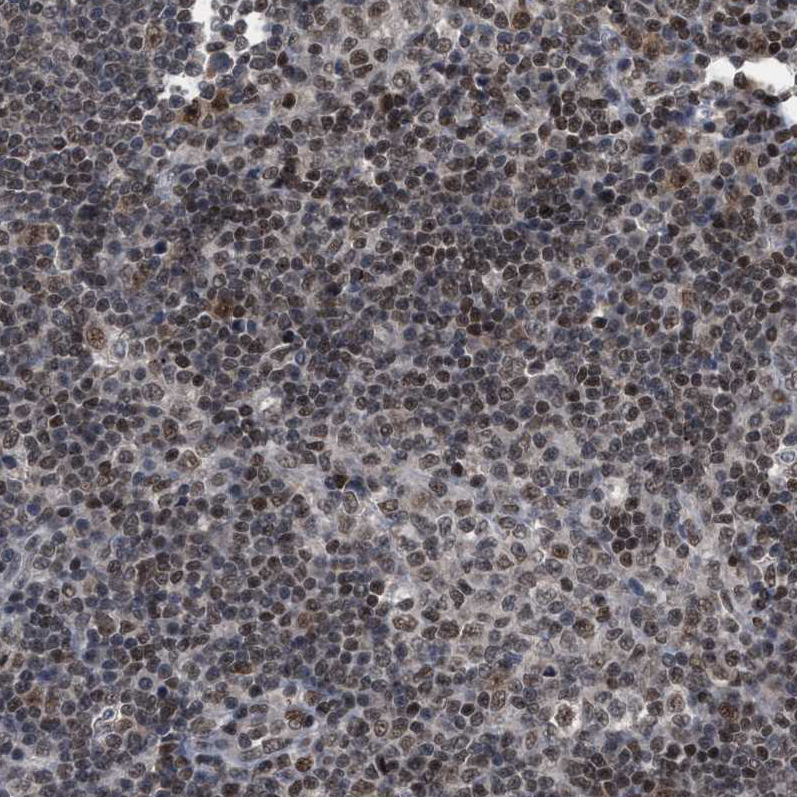
- Immunohistochemical staining of human lymph node using Anti-C9orf78 antibody HPA021232.
- Sample type
- HUMAN
- Submitted by
- Atlas Antibodies (provider)
- Main image

- Experimental details
- Immunohistochemical staining of human testis using Anti-C9orf78 antibody HPA021232.
- Sample type
- HUMAN

- Submitted by
- Atlas Antibodies (provider)
- Main image

- Experimental details
- Immunohistochemical staining of human pancreas shows weak positivity in exocrine glandular cells as expected.
- Sample type
- HUMAN

- Submitted by
- Atlas Antibodies (provider)
- Main image

- Experimental details
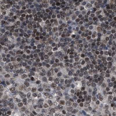
- Immunohistochemical staining of human lymph node shows strong nuclear positivity in non-germinal center cells.
- Sample type
- HUMAN
- Submitted by
- Atlas Antibodies (provider)
- Main image

- Experimental details
- Immunohistochemical staining of human Fallopian tube shows moderate nuclear positivity in glandular cells.
- Sample type
- HUMAN

- Submitted by
- Atlas Antibodies (provider)
- Main image

- Experimental details
- Immunohistochemical staining of human testis shows strong nuclear positivity in cells in seminiferous ducts.
- Sample type
- HUMAN
