Explore
 Validate
 Learn
Learn Western blot
 Immunocytochemistry
Antibody data
- Antibody Data
- Antigen structure
- References [1]
- Comments [0]
- Validations
- Western blot [4]
- Immunohistochemistry [13]
Submit
Validation data
Reference
Comment
Report error
- Product number
- NBP1-83168 - Provider product page

- Provider
- Novus Biologicals
- Proper citation
- Novus Cat#NBP1-83168, RRID:AB_11006927
- Product name
- Rabbit Polyclonal HCA59 Antibody
- Antibody type
- Polyclonal
- Description
- Immunogen affinity purified. Specificity of human, mouse, rat HCA59 antibody verified on a Protein Array containing target protein plus 383 other non-specific proteins.
- Reactivity
- Human, Mouse, Rat
- Host
- Rabbit
- Isotype
- IgG
- Vial size
- 0.1 ml
- Storage
- Store at 4C short term. Aliquot and store at -20C long term. Avoid freeze-thaw cycles.
Submitted references Contribution of antibody-based protein profiling to the human Chromosome-centric Proteome Project (C-HPP).
Fagerberg L, Oksvold P, Skogs M, Algenäs C, Lundberg E, Pontén F, Sivertsson A, Odeberg J, Klevebring D, Kampf C, Asplund A, Sjöstedt E, Al-Khalili Szigyarto C, Edqvist PH, Olsson I, Rydberg U, Hudson P, Ottosson Takanen J, Berling H, Björling L, Tegel H, Rockberg J, Nilsson P, Navani S, Jirström K, Mulder J, Schwenk JM, Zwahlen M, Hober S, Forsberg M, von Feilitzen K, Uhlén M
Journal of proteome research 2013 Jun 7;12(6):2439-48
Journal of proteome research 2013 Jun 7;12(6):2439-48
No comments: Submit comment
Supportive validation
- Submitted by
- Novus Biologicals (provider)
- Main image
- Experimental details
- Western Blot: HCA59 Antibody [NBP1-83168] - Lane 1: NIH-3T3 cell lysate (Mouse embryonic fibroblast cells). Lane 2: NBT-II cell lysate (Rat Wistar bladder tumor cells).
- Submitted by
- Novus Biologicals (provider)
- Main image
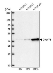
- Experimental details
- Western Blot: HCA59 Antibody [NBP1-83168] - Analysis in U2OS cells transfected with control siRNA, target specific siRNA probe #1 and #2, using anti-C9orf78 antibody. Remaining relative intensity is presented.

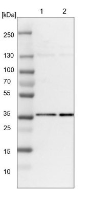
- Submitted by
- Novus Biologicals (provider)
- Main image
- Experimental details
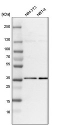
- Western Blot: HCA59 Antibody [NBP1-83168] - Analysis in mouse cell line NIH-3T3 and rat cell line NBT-II.

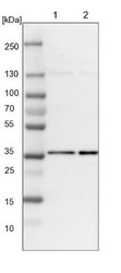
- Submitted by
- Novus Biologicals (provider)
- Main image
- Experimental details
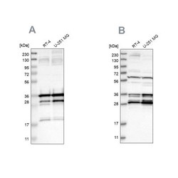
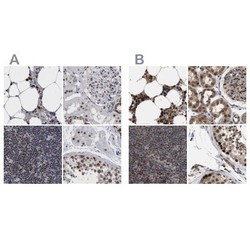
- Western Blot: HCA59 Antibody [NBP1-83168] - Analysis using Anti-C9orf78 antibody NBP1-83168 (A) shows similar pattern to independent antibody NBP1-83170 (B).

Supportive validation
- Submitted by
- Novus Biologicals (provider)
- Main image
- Experimental details
- Immunohistochemistry-Paraffin: HCA59 Antibody [NBP1-83168] - Staining of human gall bladder shows strong nuclear positivity in glandular cells.

- Submitted by
- Novus Biologicals (provider)
- Main image
- Experimental details
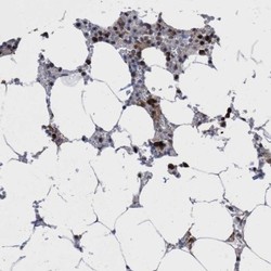
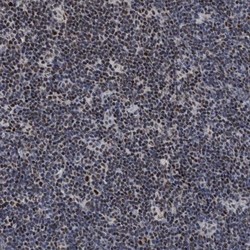
- Immunohistochemistry-Paraffin: HCA59 Antibody [NBP1-83168] - Staining of human bone marrow shows high expression.
- Submitted by
- Novus Biologicals (provider)
- Main image
- Experimental details
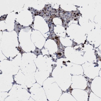
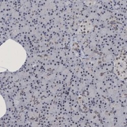
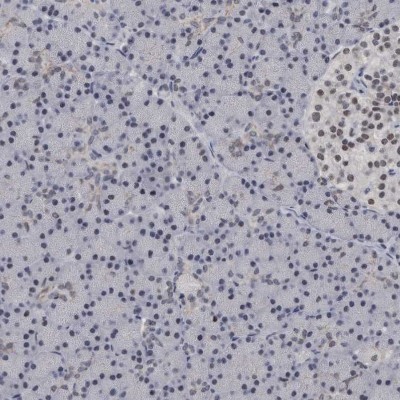
- Immunohistochemistry-Paraffin: HCA59 Antibody [NBP1-83168] - Staining of human pancreas shows low expression as expected.

- Submitted by
- Novus Biologicals (provider)
- Main image
- Experimental details
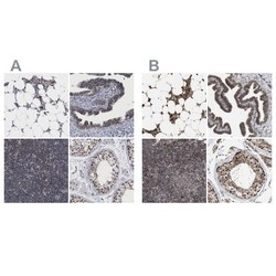
- Immunohistochemistry-Paraffin: HCA59 Antibody [NBP1-83168] - Staining of human bone marrow, kidney, lymph node and testis using Anti-C9orf78 antibody NBP1-83168 (A) shows similar protein distribution across tissues to independent antibody NBP1-83170 (B).
- Submitted by
- Novus Biologicals (provider)
- Main image
- Experimental details
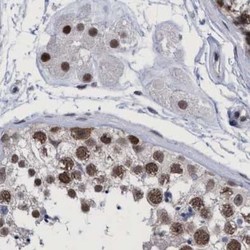
- Immunohistochemistry-Paraffin: HCA59 Antibody [NBP1-83168] - Staining of human testis.

- Submitted by
- Novus Biologicals (provider)
- Main image

- Experimental details
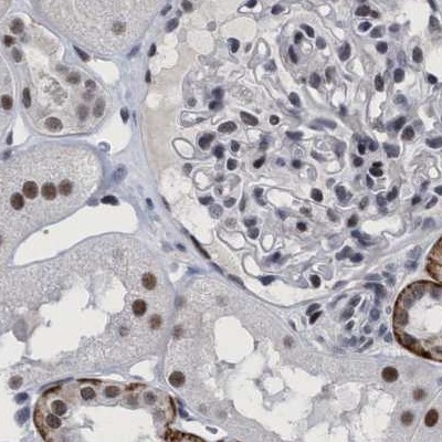
- Immunohistochemistry-Paraffin: HCA59 Antibody [NBP1-83168] - Staining of human kidney.
- Submitted by
- Novus Biologicals (provider)
- Main image
- Experimental details
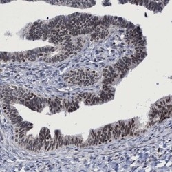
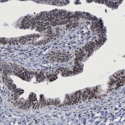
- Immunohistochemistry: HCA59 Antibody [NBP1-83168] - Staining of human Fallopian tube shows moderate nuclear positivity in glandular cells.
- Submitted by
- Novus Biologicals (provider)
- Main image
- Experimental details
- Immunohistochemistry-Paraffin: HCA59 Antibody [NBP1-83168] - Staining of human lymph node.
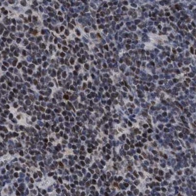
- Submitted by
- Novus Biologicals (provider)
- Main image
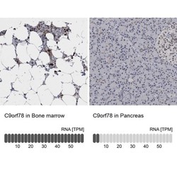
- Experimental details
- Immunohistochemistry-Paraffin: HCA59 Antibody [NBP1-83168] - Analysis in human bone marrow and pancreas tissues. Corresponding HCA59 RNA-seq data are presented for the same tissues.
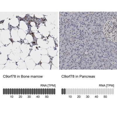
- Submitted by
- Novus Biologicals (provider)
- Main image
- Experimental details
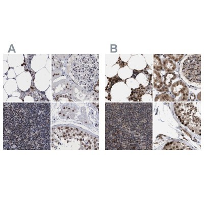
- Immunohistochemistry-Paraffin: HCA59 Antibody [NBP1-83168] - Staining of human bone marrow, fallopian tube, lymph node and testis using Anti-HCA59 antibody NBP1-83168 (A) shows similar protein distribution across tissues to independent antibody NBP1-83170 (B).

- Submitted by
- Novus Biologicals (provider)
- Main image
- Experimental details
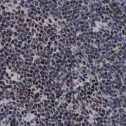
- Immunohistochemistry-Paraffin: HCA59 Antibody [NBP1-83168] - Staining of human lymph node shows strong nuclear positivity in non-germinal center cells.

- Submitted by
- Novus Biologicals (provider)
- Main image

- Experimental details
- Immunohistochemistry-Paraffin: HCA59 Antibody [NBP1-83168] - Staining of human pancreas shows very weak positivity in exocrine glandular cells as expected.
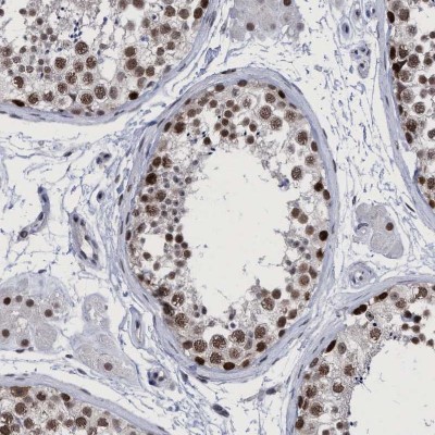
- Submitted by
- Novus Biologicals (provider)
- Main image
- Experimental details
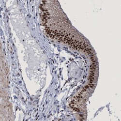
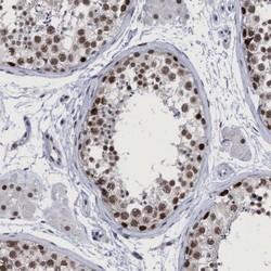
- Immunohistochemistry-Paraffin: HCA59 Antibody [NBP1-83168] - Staining of human testis shows strong nuclear positivity in cells in seminiferous ducts.