Explore
 Validate
 Learn
Learn Immunohistochemistry
Antibody data
- Antibody Data
- Antigen structure
- References [2]
- Comments [0]
- Validations
- Immunohistochemistry [7]
Submit
Validation data
Reference
Comment
Report error
- Product number
- HPA021004 - Provider product page

- Provider
- Atlas Antibodies
- Proper citation
- Atlas Antibodies Cat#HPA021004, RRID:AB_1854232
- Product name
- Anti-MYBPC1
- Antibody type
- Polyclonal
- Reactivity
- Human
- Host
- Rabbit
- Conjugate
- Unconjugated
- Antigen sequence
DWTLVETPPGEEQAKQNANSQLSILFIEKPQGGTV
KVGEDITFIAKVKAEDLLRKPTIKWFKGKWMDLAS
KAGKHLQLKETFERHSRVYTFEMQIIKAKDNFAGN
YRCEVTYKDKFDSCSFDLEVHESTGTTPN- Isotype
- IgG
- Vial size
- 100 µl
- Storage
- Store at +4°C for short term storage. Long time storage is recommended at -20°C.
Submitted references Immunofluorescence and fluorescent-protein tagging show high correlation for protein localization in mammalian cells
Expression profiles of muscle disease-associated genes and their isoforms during differentiation of cultured human skeletal muscle cells.
Stadler C, Rexhepaj E, Singan V, Murphy R, Pepperkok R, Uhlén M, Simpson J, Lundberg E
Nature Methods 2013 February;10(4):315-323
Nature Methods 2013 February;10(4):315-323
Expression profiles of muscle disease-associated genes and their isoforms during differentiation of cultured human skeletal muscle cells.
Abdul-Hussein S, van der Ven PF, Tajsharghi H
BMC musculoskeletal disorders 2012 Dec 29;13:262
BMC musculoskeletal disorders 2012 Dec 29;13:262
No comments: Submit comment
Enhanced validation
Enhanced validation
Supportive validation
- Submitted by
- Atlas Antibodies (provider)
- Enhanced method
- Orthogonal validation
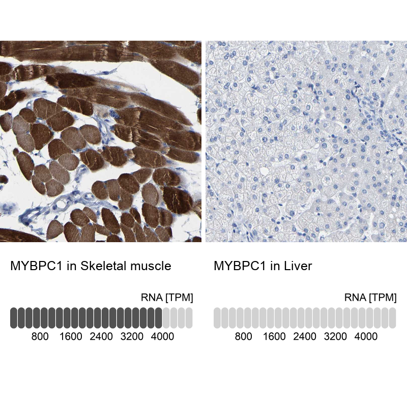
- Main image

- Experimental details
- Immunohistochemistry analysis in human skeletal muscle and liver tissues using Anti-MYBPC1 antibody. Corresponding MYBPC1 RNA-seq data are presented for the same tissues.
- Sample type
- HUMAN
Enhanced validation
- Submitted by
- Atlas Antibodies (provider)
- Enhanced method
- Independent antibody validation
- Main image
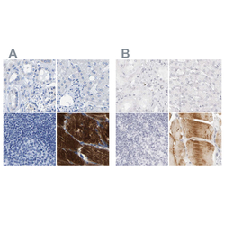
- Experimental details
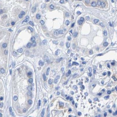
- Immunohistochemical staining of human kidney, liver, lymph node and skeletal muscle using Anti-MYBPC1 antibody HPA021004 (A) shows similar protein distribution across tissues to independent antibody HPA027614 (B).

Supportive validation
- Submitted by
- Atlas Antibodies (provider)
- Main image
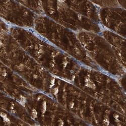
- Experimental details
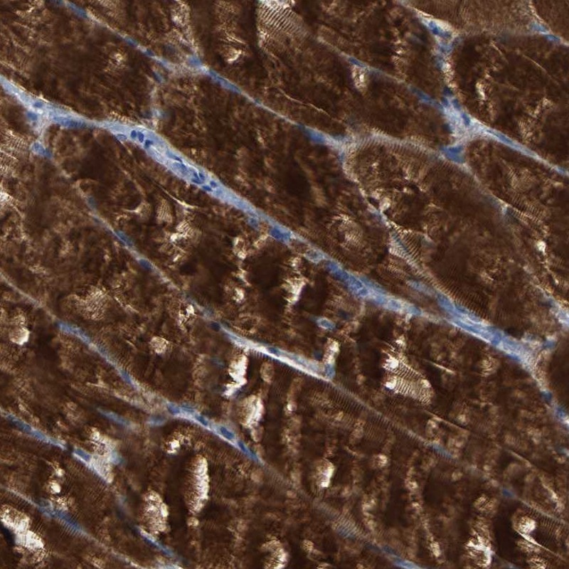
- Immunohistochemical staining of human skeletal muscle shows strong cytoplasmic positivity in myocytes.
- Submitted by
- Atlas Antibodies (provider)
- Main image
- Experimental details
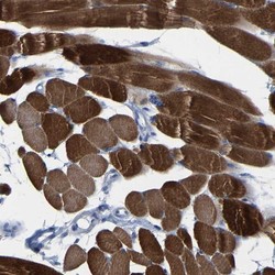
- Immunohistochemical staining of human skeletal muscle shows high expression.
- Sample type
- HUMAN

- Submitted by
- Atlas Antibodies (provider)
- Main image
- Experimental details
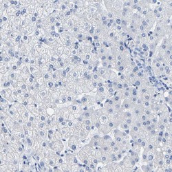
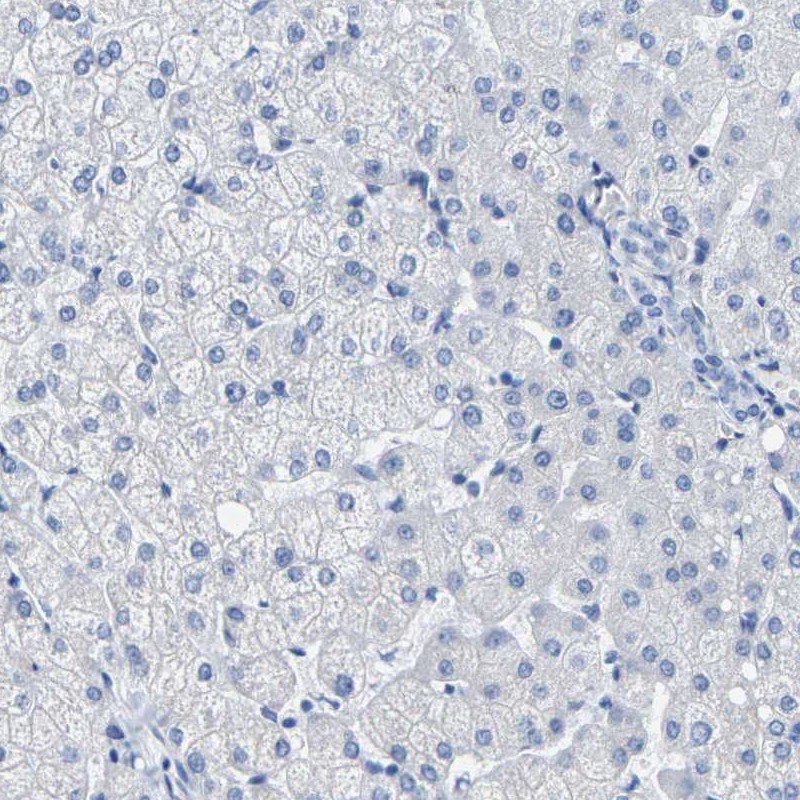
- Immunohistochemical staining of human liver shows low expression as expected.
- Sample type
- HUMAN
- Submitted by
- Atlas Antibodies (provider)
- Main image
- Experimental details
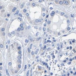
- Immunohistochemical staining of human kidney using Anti-MYBPC1 antibody HPA021004.
- Sample type
- HUMAN
- Submitted by
- Atlas Antibodies (provider)
- Main image
- Experimental details
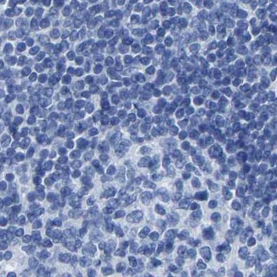
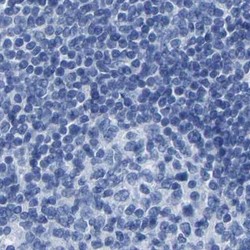
- Immunohistochemical staining of human lymph node using Anti-MYBPC1 antibody HPA021004.
- Sample type
- HUMAN