 Explore
 Validate
 Learn
Learn Western blot
Antibody data
- Antibody Data
- Antigen structure
- References [0]
- Comments [0]
- Validations
- Western blot [4]
- Immunocytochemistry [1]
- Immunoprecipitation [1]
- Immunohistochemistry [3]
Submit
Validation data
Reference
Comment
Report error
- Product number
- GTX104531 - Provider product page

- Provider
- GeneTex
- Proper citation
- GeneTex Cat#GTX104531, RRID:AB_1950805
- Product name
- LDB1 antibody [N2C3]
- Antibody type
- Polyclonal
- Reactivity
- Human, Mouse, Rat
- Host
- Rabbit
No comments: Submit comment
Supportive validation
- Submitted by
- GeneTex (provider)
- Main image
- Experimental details
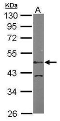
- Sample (50 ?g of whole cell lysate) A: Mouse brain 10% SDS PAGE GTX104531 diluted at 1:1000 The HRP-conjugated anti-rabbit IgG antibody (GTX213110-01) was used to detect the primary antibody.

- Submitted by
- GeneTex (provider)
- Main image
- Experimental details
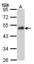
- Sample (30 ug of whole cell lysate) A: Raji 10% SDS PAGE GTX104531 diluted at 1:1000
- Validation comment
- WB

- Submitted by
- GeneTex (provider)
- Main image
- Experimental details
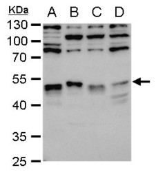
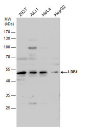
- LDB1 antibody [N2C3] detects LDB1 protein by western blot analysis.A. 30 ?g 293T whole cell extract B. 30 ?g A431 whole cell extract C. 30 ?g HeLa whole cell extract D. 30 ?g HepG2 whole cell extract10 % SDS-PAGELDB1 antibody [N2C3] (GTX104531) dilution: 1:1000
- Validation comment
- WB
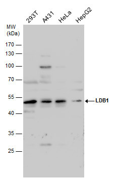
- Submitted by
- GeneTex (provider)
- Main image
- Experimental details
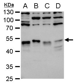
- LDB1 antibody detects LDB1 protein by western blot analysis. Various whole cell extracts (30 ?g) were separated by 10% SDS-PAGE, and the membrane was blotted with LDB1 antibody (GTX104531) diluted by 1:1000. The HRP-conjugated anti-rabbit IgG antibody (GTX213110-01) was used to detect the primary antibody.
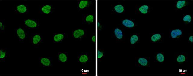
Supportive validation
- Submitted by
- GeneTex (provider)
- Main image

- Experimental details
- LDB1 antibody [N2C3] detects LDB1 protein at nucleus by immunofluorescent analysis.Sample: HeLa cells were fixed in 4% paraformaldehyde at RT for 15 min.Green: LDB1 protein stained by LDB1 antibody [N2C3] (GTX104531) diluted at 1:500.Blue: Hoechst 33342 staining.Scale bar = 10 £gm.
Supportive validation
- Submitted by
- GeneTex (provider)
- Main image
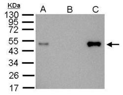
- Experimental details
- LDB1antibody immunoprecipitates LDB1 protein in IP experiments.IP Sample: 1000 ?g 293T whole cell lysate/extract A. 20 £gg 293T whole cell lysate/extractB. Control with 2.5 £gg of preimmune rabbit IgGC. Immunoprecipitation of LDB1 protein by 2.5 £gg of LDB1 antibody (GTX104531)12% SDS-PAGE The immunoprecipitated LDB1 protein was detected by LDB1antibody (GTX104531) diluted at 1:1000. EasyBlot anti-rabbit IgG (GTX221666-01) was used as a secondary reagent.

Supportive validation
- Submitted by
- GeneTex (provider)
- Main image
- Experimental details
- Immunohistochemical analysis of paraffin-embedded U87 Xenograft, using LDB1(GTX104531) antibody at 1:100 dilution.

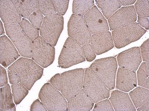
- Submitted by
- GeneTex (provider)
- Main image

- Experimental details
- LDB1 antibody [N2C3] detects LDB1 protein at nucleus on mouse muscle by immunohistochemical analysis. Sample: Paraffin-embedded mouse muscle. LDB1 antibody [N2C3] (GTX104531) dilution: 1:500.
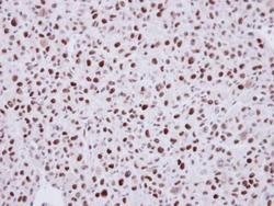
- Submitted by
- GeneTex (provider)
- Main image

- Experimental details
- LDB1 antibody [N2C3] detects LDB1 protein at nucleus on rat middle brain by immunohistochemical analysis. Sample: Paraffin-embedded rat middle brain. LDB1 antibody [N2C3] (GTX104531) dilution: 1:500.
