 Explore
 Validate
 Learn
Learn Western blot
 Immunoprecipitation
Antibody data
- Antibody Data
- Antigen structure
- References [0]
- Comments [0]
- Validations
- Western blot [8]
- Immunocytochemistry [2]
- Immunohistochemistry [2]
- Other assay [1]
Submit
Validation data
Reference
Comment
Report error
- Product number
- 14805-1-AP - Provider product page

- Provider
- Invitrogen Antibodies
- Product name
- EXOSC2 Polyclonal Antibody
- Antibody type
- Polyclonal
- Antigen
- Other
- Description
- Immunogen sequence: MAMEMRLPV ARKPLSERLG RDTKKHLVVP GDTITTDTGF MRGHGTYMGE EKLIASVAGS VERVNKLICV KALKTRYIGE VGDIVVGRIT EVQQKRWKVE TNSRLDSVLL LSSMNLPGGE LRRRSAEDEL AMRGFLQEGD LISAEVQAVF SDGAVSLHTR SLKYGKLGQG VLVQVSPSLV KRQKTHFHDL PCGASVILGN NGFIWIYPTP EHKEEEAGGF IANLEPVSLA DREVISRLRN CIISLVTQRM MLYDTSILYC YEASLPHQIK DILKPEIMEE IVMETRQRLL EQEG (1-293 aa encoded by BC000747)
- Reactivity
- Human
- Host
- Rabbit
- Isotype
- IgG
- Vial size
- 150 µL
- Concentration
- 0.21 mg/mL
- Storage
- -20°C
No comments: Submit comment
Supportive validation
- Submitted by
- Invitrogen Antibodies (provider)
- Main image

- Experimental details
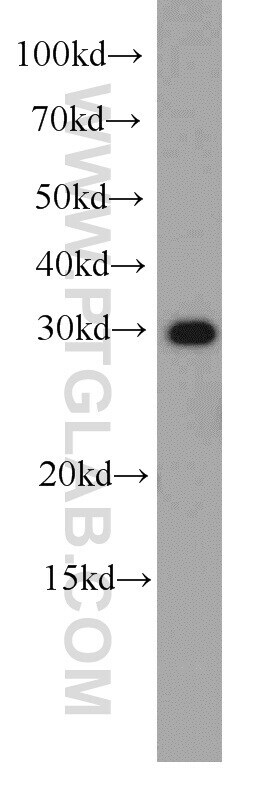
- HeLa cells were subjected to SDS PAGE followed by western blot with 14805-1-AP (EXOSC2 antibody) at dilution of 1:1000 incubated at room temperature for 1.5 hours.

- Submitted by
- Invitrogen Antibodies (provider)
- Main image

- Experimental details
- Jurkat cells were subjected to SDS PAGE followed by western blot with 14805-1-AP (EXOSC2 antibody) at dilution of 1:1000 incubated at room temperature for 1.5 hours.
- Submitted by
- Invitrogen Antibodies (provider)
- Main image

- Experimental details
- HeLa cells were subjected to SDS PAGE followed by western blot with 14805-1-AP (EXOSC2 antibody) at dilution of 1:1000 incubated at room temperature for 1.5 hours.

- Submitted by
- Invitrogen Antibodies (provider)
- Main image

- Experimental details
- MCF7 cells were subjected to SDS PAGE followed by western blot with 14805-1-AP (EXOSC2 antibody) at dilution of 1:1000 incubated at room temperature for 1.5 hours.

- Submitted by
- Invitrogen Antibodies (provider)
- Main image

- Experimental details
- HeLa cells were subjected to SDS PAGE followed by western blot with 14805-1-AP (EXOSC2 Antibody) at dilution of 1:1000 incubated at room temperature for 1.5 hours.

- Submitted by
- Invitrogen Antibodies (provider)
- Main image

- Experimental details
- HepG2 cells were subjected to SDS PAGE followed by western blot with 14805-1-AP (EXOSC2 Antibody) at dilution of 1:1000 incubated at room temperature for 1.5 hours.

- Submitted by
- Invitrogen Antibodies (provider)
- Main image

- Experimental details
- HeLa cells were subjected to SDS PAGE followed by western blot with 14805-1-AP (EXOSC2 antibody) at dilution of 1:500 incubated at room temperature for 1.5 hours.
- Submitted by
- Invitrogen Antibodies (provider)
- Main image

- Experimental details
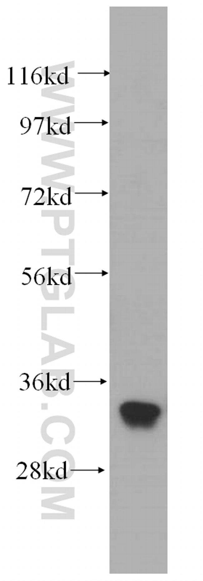
- WB result of EXOSC2 antibody (14805-1-AP; 1:10000; incubated at room temperature for 1.5 hours) with sh-Control and sh-EXOSC2 transfected HEK-293 cells.

Supportive validation
- Submitted by
- Invitrogen Antibodies (provider)
- Main image

- Experimental details
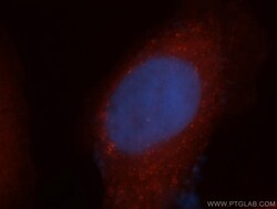
- Immunofluorescent analysis of MCF-7 cells, using EXOSC2 antibody 14805-1-AP at 1:50 dilution and Rhodamine-labeled goat anti-rabbit IGG (red). Blue pseudocolor = DAPI (fluorescent DNA dye).

- Submitted by
- Invitrogen Antibodies (provider)
- Main image
- Experimental details
- Immunofluorescent analysis of MCF-7 cells, using EXOSC2 antibody 14805-1-AP at 1:50 dilution and Rhodamine-labeled goat anti-rabbit IGG (red). Blue pseudocolor = DAPI (fluorescent DNA dye).

Supportive validation
- Submitted by
- Invitrogen Antibodies (provider)
- Main image

- Experimental details
- Immunohistochemistry of paraffin-embedded human skin cancer using 14805-1-AP (EXOSC2 antibody) at dilution of 1:100 (under 10x lens).

- Submitted by
- Invitrogen Antibodies (provider)
- Main image

- Experimental details
- Immunohistochemistry of paraffin-embedded human skin cancer using 14805-1-AP (EXOSC2 antibody) at dilution of 1:100 (under 40x lens).

Supportive validation
- Submitted by
- Invitrogen Antibodies (provider)
- Main image

- Experimental details
- IP result of anti-EXOSC2 (IP:14805-1-AP, 4ug; Detection:14805-1-AP 1:1000) with HeLa cells lysate 1080ug.
