Explore
 Validate
 Learn
Learn ELISA
ELISA Flow cytometry
Antibody data
- Antibody Data
- Antigen structure
- References [1]
- Comments [0]
- Validations
- ELISA [1]
- Immunohistochemistry [1]
Submit
Validation data
Reference
Comment
Report error
- Product number
- MAB9081 - Provider product page

- Provider
- Novus Biologicals
- Product name
- Mouse Monoclonal MMP-8 Antibody
- Antibody type
- Monoclonal
- Description
- Protein A or G purified from ascites. Detects both pro and active forms of human MMP-8 in Western blots. In Western blots, no cross-reactivity with recombinant human (rh) MMP-1, -2, -3, -7, -9, -10, -12, or -13 is observed.
- Reactivity
- Human
- Host
- Mouse
- Conjugate
- Unconjugated
- Isotype
- IgG
- Vial size
- 500 ug
- Storage
- Use a manual defrost freezer and avoid repeated freeze-thaw cycles. 12 months from date of receipt, -20 to -70 degreesC as supplied. 1 month, 2 to 8 degreesC under sterile conditions after reconstitution. 6 months, -20 to -70 degreesC under sterile conditions after reconstitution.
Submitted references Matrix metalloproteinase-8 is expressed in human chorion during labor.
Arechavaleta-Velasco F, Marciano D, Díaz-Cueto L, Parry S
American journal of obstetrics and gynecology 2004 Mar;190(3):843-50
American journal of obstetrics and gynecology 2004 Mar;190(3):843-50
No comments: Submit comment
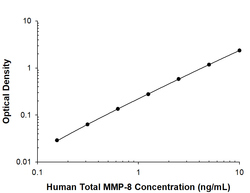
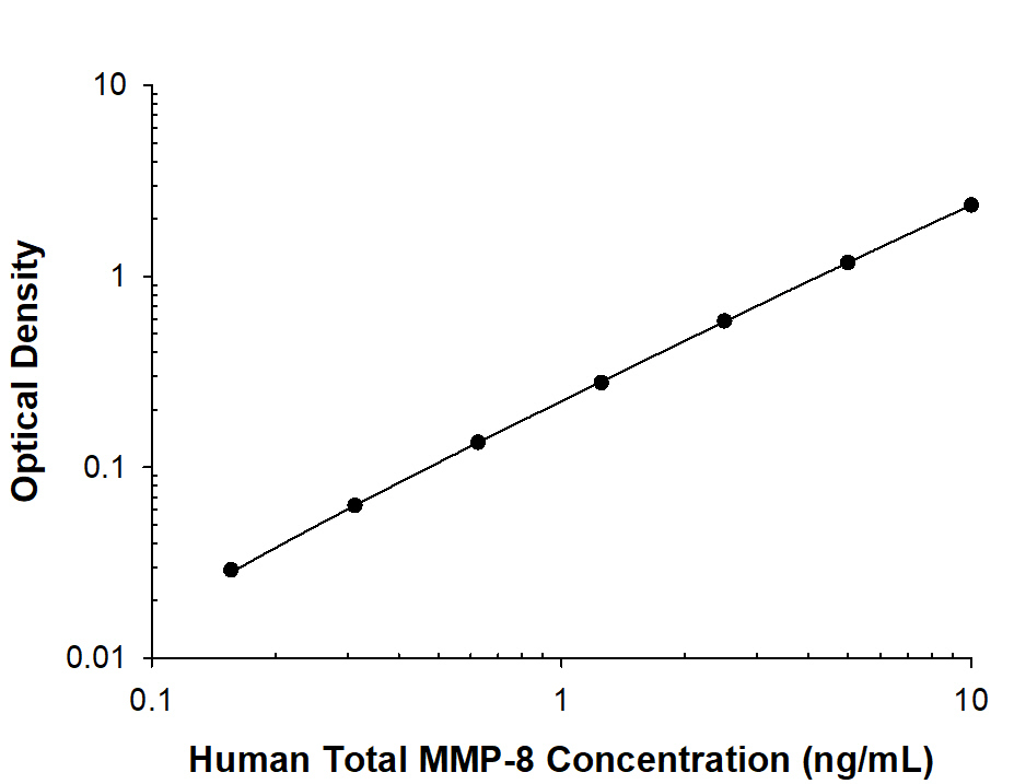
Supportive validation
- Submitted by
- Novus Biologicals (provider)
- Main image
- Experimental details
- Human MMP-8 ELISA Standard Curve.
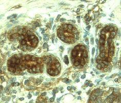
Supportive validation
- Submitted by
- Novus Biologicals (provider)
- Main image

- Experimental details
- MMP-8 in Human Breast Cancer Tissue. MMP-8 was detected in immersion fixed paraffin-embedded sections of human breast cancer tissue using 8 µg/mL Mouse Anti-Human MMP-8 Monoclonal Antibody (Catalog # MAB9081) overnight at 4 °C. Tissue was stained with the Anti-Mouse HRP-DAB Cell & Tissue Staining Kit (brown; Catalog # CTS002) and counterstained with hematoxylin (blue). Specific labeling was localized to epithelial cells in terminal ductules (round) and intralobular duct (elongated) composing glandular lobules. View our protocol for Chromogenic IHC Staining of Paraffin-embedded Tissue Sections.