Explore
 Validate
 Learn
Learn Western blot
Antibody data
- Antibody Data
- Antigen structure
- References [0]
- Comments [0]
- Validations
- Western blot [9]
- Immunocytochemistry [1]
- Immunohistochemistry [1]
Submit
Validation data
Reference
Comment
Report error
- Product number
- PA5-77868 - Provider product page

- Provider
- Invitrogen Antibodies
- Product name
- ASCL1 Polyclonal Antibody
- Antibody type
- Polyclonal
- Antigen
- Recombinant full-length protein
- Description
- Positive Control: U87-MG, SK-N-SH, IMR32, SK-N-AS, SH-SY5Y, SH-SY5Y nuclear extract, PC-12, Rat2 Predicted Reactivity: Mouse (84%) Store product as a concentrated solution. Centrifuge briefly prior to opening the vial.
- Reactivity
- Human, Mouse, Rat
- Host
- Rabbit
- Isotype
- IgG
- Vial size
- 100 µL
- Concentration
- 1.42 mg/mL
- Storage
- Store at 4°C short term. For long term storage, store at -20°C, avoiding freeze/thaw cycles.
No comments: Submit comment
Supportive validation
- Submitted by
- Invitrogen Antibodies (provider)
- Main image
- Experimental details
- Western blot analysis of ASCL1 in whole cell lysate using 30 μg of protein. Samples were separated with 15 % SDS-PAGE and incubated with ASCL1 polyclonal antibody (Product # PA5-77868) using a dilution of 1:1000.

- Submitted by
- Invitrogen Antibodies (provider)
- Main image
- Experimental details
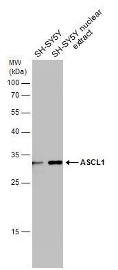
- Western blot analysis of ASCL1 in SH-SY5Y cells using 30 µg of protein. Samples were separated with 12% SDS-PAGE and incubated with ASCL1 polyclonal antibody (Product # PA5-77868) using a dilution of 1:1000.
- Submitted by
- Invitrogen Antibodies (provider)
- Main image
- Experimental details
- Western blot analysis of ASCL1 in whole cell lysate using 30 µg of protein. Samples were separated with 12% SDS-PAGE and incubated with ASCL1 polyclonal antibody (Product # PA5-77868) using a dilution of 1:1000.
- Submitted by
- Invitrogen Antibodies (provider)
- Main image
- Experimental details
- Western blot analysis of ASCL1 in whole cell lysate using 30 µg of protein. Samples were separated with 12% SDS-PAGE and incubated with ASCL1 polyclonal antibody (Product # PA5-77868) using a dilution of 1:1000.
- Submitted by
- Invitrogen Antibodies (provider)
- Main image
- Experimental details
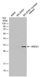
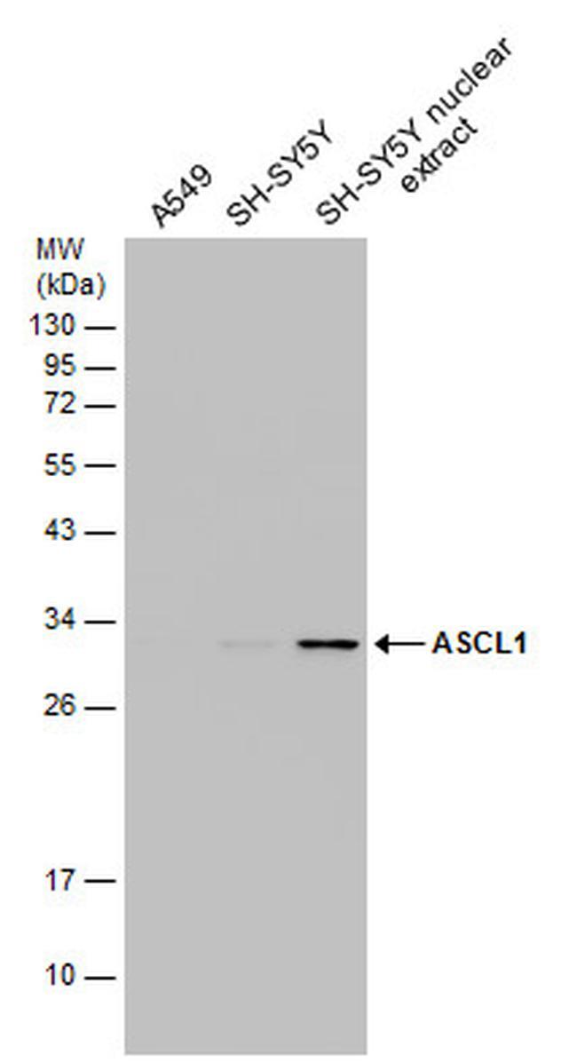
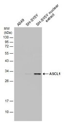
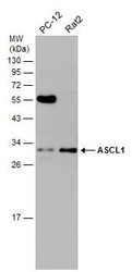
- Western Blot using ASCL1 Polyclonal Antibody (Product # PA5-77868). Various whole cell and SH-SY5Y nuclear extracts (30 µg) were separated by 12% SDS-PAGE, and the membrane was blotted with ASCL1 Polyclonal Antibody (Product # PA5-77868) diluted at 1:1,000.
- Submitted by
- Invitrogen Antibodies (provider)
- Main image
- Experimental details
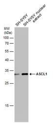
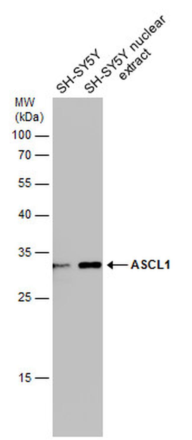
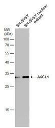
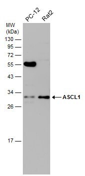
- Western Blot using ASCL1 Polyclonal Antibody (Product # PA5-77868). SH-SY5Y whole cell and nuclear extracts (30 µg) were separated by 12% SDS-PAGE, and the membrane was blotted with ASCL1 Polyclonal Antibody (Product # PA5-77868) diluted at 1:1,000.
- Submitted by
- Invitrogen Antibodies (provider)
- Main image
- Experimental details
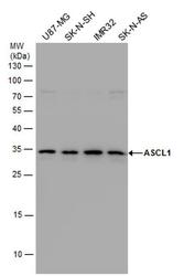
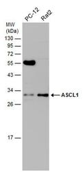
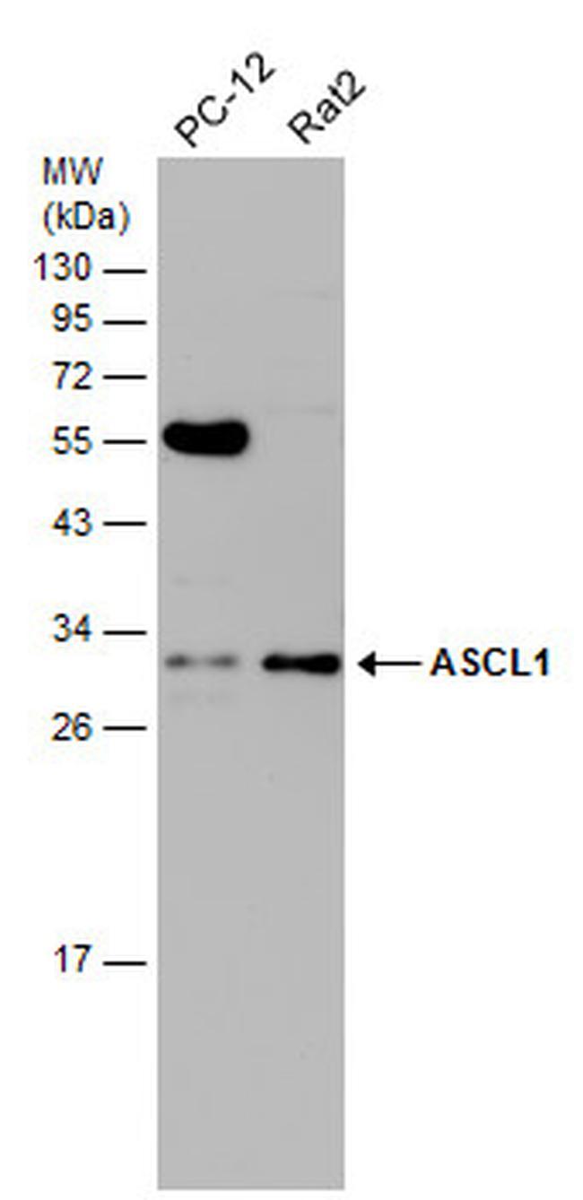
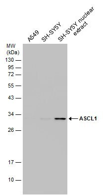
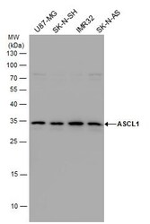
- Western Blot using ASCL1 Polyclonal Antibody (Product # PA5-77868). Various whole cell extracts (30 µg) were separated by 12% SDS-PAGE, and the membrane was blotted with ASCL1 Polyclonal Antibody (Product # PA5-77868) diluted at 1:1,000.
- Submitted by
- Invitrogen Antibodies (provider)
- Main image
- Experimental details
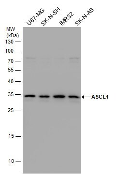
- ASCL1 Polyclonal Antibody detects ASCL1 protein by western blot analysis. Various whole cell extracts (30 µg) were separated by 15 % SDS-PAGE, and blotted with ASCL1 Polyclonal Antibody (Product # PA5-77868) diluted by 1:1,000.
- Submitted by
- Invitrogen Antibodies (provider)
- Main image
- Experimental details
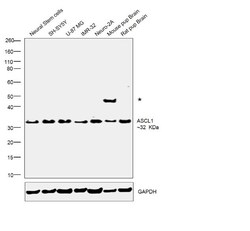
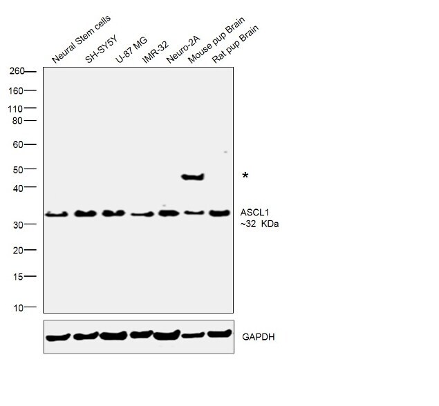
- Western blot was performed using Anti-ASCL1 Polyclonal Antibody (Product # PA5-77868) and 32 kDa band was observed corresponding to ASCL1 was observed across the cell lines tested along with an uncharacterized band (*) in Mouse pup brain. Modified Whole cell extracts (30ug lysate) (1% SDS) of Neural Stem cells (Lane 1), SH-SY5Y (Lane 2), U-87 MG (Lane 3), IMR-32 (Lane 4), Neuro-2A (Lane 5), tissue extracts of Mouse pup brain (Lane 6) and Rat pup brain (Lane 7) were electrophoresed using Novex® NuPAGE® 4-12 % Bis-Tris gel (Product # NP0322BOX). Resolved proteins were then transferred onto a nitrocellulose membrane (Product # IB23001) by iBlot® 2 Dry Blotting System (Product # IB21001). The blot was probed with the primary antibody (1:1000 dilution) and detected by chemiluminescence with Goat anti-Rabbit IgG (H+L) Superclonal™ Recombinant Secondary Antibody, HRP (Product # A27036, 1:4000 dilution) using the iBright FL 1000 (Product # A32752). Chemiluminescent detection was performed using Novex® ECL Chemiluminescent Substrate Reagent Kit (Product # WP20005)..
Supportive validation
- Submitted by
- Invitrogen Antibodies (provider)
- Main image
- Experimental details
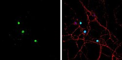
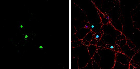
- Immunocytochemistry-Immunofluorescence analysis of ASCL1 was performed in DIV9 rat E18 primary hippocampal neuron cells fixed in 4% paraformaldehyde at RT for 15 min. Green: ASCL1 Polyclonal Antibody (Product # PA5-77868) diluted at 1:500. Red: beta Tubulin 3/ Tuj1, stained by beta Tubulin 3/ Tuj1 antibody. Blue: Fluoroshield with DAPI.
Supportive validation
- Submitted by
- Invitrogen Antibodies (provider)
- Main image
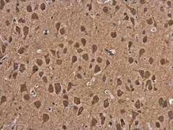
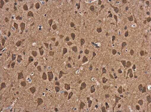
- Experimental details
- ASCL1 Polyclonal Antibody detects ASCL1 protein at cytoplasm in rat brain by immunohistochemical analysis. Sample: Paraffin-embedded rat brain. ASCL1 Polyclonal Antibody (Product # PA5-77868) diluted at 1:500. Antigen Retrieval: Citrate buffer, pH 6.0, 15 min.