 Explore
 Validate
 Learn
Learn Western blot
Antibody data
- Antibody Data
- Antigen structure
- References [30]
- Comments [0]
- Validations
- Western blot [10]
- Immunocytochemistry [1]
- Immunoprecipitation [1]
- Immunohistochemistry [9]
Submit
Validation data
Reference
Comment
Report error
- Product number
- GTX128145 - Provider product page

- Provider
- GeneTex
- Proper citation
- GeneTex Cat#GTX128145, RRID:AB_2687562
- Product name
- ATR (phospho Thr1989) antibody
- Antibody type
- Polyclonal
- Reactivity
- Human, Mouse
- Host
- Rabbit
Submitted references Preclinical study using androgen receptor (AR) degradation enhancer to increase radiotherapy efficacy via targeting radiation-increased AR to better suppress prostate cancer progression.
pRAD50: a novel and clinically applicable pharmacodynamic biomarker of both ATM and ATR inhibition identified using mass spectrometry and immunohistochemistry.
Overexpression of the base excision repair NTHL1 glycosylase causes genomic instability and early cellular hallmarks of cancer.
TP53 Haploinsufficiency Rescues Emergency Granulopoiesis in FANCC-/- Mice.
Human Parvovirus B19 Utilizes Cellular DNA Replication Machinery for Viral DNA Replication.
Loss of Wilms tumor 1 protein is a marker for apoptosis in response to replicative stress in leukemic cells.
BRD4 facilitates replication stress-induced DNA damage response.
Induced Telomere Damage to Treat Telomerase Expressing Therapy-Resistant Pediatric Brain Tumors.
hDNA2 nuclease/helicase promotes centromeric DNA replication and genome stability.
Caspase-10: a molecular switch from cell-autonomous apoptosis to communal cell death in response to chemotherapeutic drug treatment.
PIDD mediates the association of DNA-PKcs and ATR at stalled replication forks to facilitate the ATR signaling pathway.
HDAC1 and HDAC2 integrate checkpoint kinase phosphorylation and cell fate through the phosphatase-2A subunit PR130.
The ATR Inhibitor AZD6738 Synergizes with Gemcitabine In Vitro and In Vivo to Induce Pancreatic Ductal Adenocarcinoma Regression.
MAST1 Drives Cisplatin Resistance in Human Cancers by Rewiring cRaf-Independent MEK Activation.
The G2 checkpoint inhibitor CBP-93872 increases the sensitivity of colorectal and pancreatic cancer cells to chemotherapy.
The endonuclease EEPD1 mediates synthetic lethality in RAD52-depleted BRCA1 mutant breast cancer cells.
Phosphorylated STAT5 directly facilitates parvovirus B19 DNA replication in human erythroid progenitors through interaction with the MCM complex.
Identification of the proteome complement of humanTLK1 reveals it binds and phosphorylates NEK1 regulating its activity.
Parvovirus B19 NS1 protein induces cell cycle arrest at G2-phase by activating the ATR-CDC25C-CDK1 pathway.
A Class of Environmental and Endogenous Toxins Induces BRCA2 Haploinsufficiency and Genome Instability.
DNA damage-induced ATM- and Rad-3-related (ATR) kinase activation in non-replicating cells is regulated by the XPB subunit of transcription factor IIH (TFIIH).
Reevaluation of ATR signaling in primary resting chronic lymphocytic leukemia cells: evidence for pro-survival or pro-apoptotic function.
BET bromodomain inhibitors synergize with ATR inhibitors in melanoma.
RAD18, WRNIP1 and ATMIN promote ATM signalling in response to replication stress.
Replication of an Autonomous Human Parvovirus in Non-dividing Human Airway Epithelium Is Facilitated through the DNA Damage and Repair Pathways.
ATR inhibitors as a synthetic lethal therapy for tumours deficient in ARID1A.
Function of high-mobility group A proteins in the DNA damage signaling for the induction of apoptosis.
Trovafloxacin-induced replication stress sensitizes HepG2 cells to tumor necrosis factor-alpha-induced cytotoxicity mediated by extracellular signal-regulated kinase and ataxia telangiectasia and Rad3-related.
EEPD1 Rescues Stressed Replication Forks and Maintains Genome Stability by Promoting End Resection and Homologous Recombination Repair.
RPA inhibition increases replication stress and suppresses tumor growth.
Chou FJ, Chen Y, Chen D, Niu Y, Li G, Keng P, Yeh S, Chang C
EBioMedicine 2019 Feb;40:504-516
EBioMedicine 2019 Feb;40:504-516
pRAD50: a novel and clinically applicable pharmacodynamic biomarker of both ATM and ATR inhibition identified using mass spectrometry and immunohistochemistry.
Jones GN, Rooney C, Griffin N, Roudier M, Young LA, Garcia-Trinidad A, Hughes GD, Whiteaker JR, Wilson Z, Odedra R, Zhao L, Ivey RG, Howat WJ, Harrington EA, Barrett JC, Ramos-Montoya A, Lau A, Paulovich AG, Cadogan EB, Pierce AJ
British journal of cancer 2018 Nov;119(10):1233-1243
British journal of cancer 2018 Nov;119(10):1233-1243
Overexpression of the base excision repair NTHL1 glycosylase causes genomic instability and early cellular hallmarks of cancer.
Limpose KL, Trego KS, Li Z, Leung SW, Sarker AH, Shah JA, Ramalingam SS, Werner EM, Dynan WS, Cooper PK, Corbett AH, Doetsch PW
Nucleic acids research 2018 May 18;46(9):4515-4532
Nucleic acids research 2018 May 18;46(9):4515-4532
TP53 Haploinsufficiency Rescues Emergency Granulopoiesis in FANCC-/- Mice.
Hu L, Huang W, Bei L, Broglie L, Eklund EA
Journal of immunology (Baltimore, Md. : 1950) 2018 Mar 15;200(6):2129-2139
Journal of immunology (Baltimore, Md. : 1950) 2018 Mar 15;200(6):2129-2139
Human Parvovirus B19 Utilizes Cellular DNA Replication Machinery for Viral DNA Replication.
Zou W, Wang Z, Xiong M, Chen AY, Xu P, Ganaie SS, Badawi Y, Kleiboeker S, Nishimune H, Ye SQ, Qiu J
Journal of virology 2018 Mar 1;92(5)
Journal of virology 2018 Mar 1;92(5)
Loss of Wilms tumor 1 protein is a marker for apoptosis in response to replicative stress in leukemic cells.
Pons M, Reichardt CM, Hennig D, Nathan A, Kiweler N, Stocking C, Wichmann C, Christmann M, Butter F, Reichardt S, Schneider G, Heinzel T, Englert C, Hartkamp J, Krämer OH, Mahendrarajah N
Archives of toxicology 2018 Jun;92(6):2119-2135
Archives of toxicology 2018 Jun;92(6):2119-2135
BRD4 facilitates replication stress-induced DNA damage response.
Zhang J, Dulak AM, Hattersley MM, Willis BS, Nikkilä J, Wang A, Lau A, Reimer C, Zinda M, Fawell SE, Mills GB, Chen H
Oncogene 2018 Jul;37(28):3763-3777
Oncogene 2018 Jul;37(28):3763-3777
Induced Telomere Damage to Treat Telomerase Expressing Therapy-Resistant Pediatric Brain Tumors.
Sengupta S, Sobo M, Lee K, Senthil Kumar S, White AR, Mender I, Fuller C, Chow LML, Fouladi M, Shay JW, Drissi R
Molecular cancer therapeutics 2018 Jul;17(7):1504-1514
Molecular cancer therapeutics 2018 Jul;17(7):1504-1514
hDNA2 nuclease/helicase promotes centromeric DNA replication and genome stability.
Li Z, Liu B, Jin W, Wu X, Zhou M, Liu VZ, Goel A, Shen Z, Zheng L, Shen B
The EMBO journal 2018 Jul 13;37(14)
The EMBO journal 2018 Jul 13;37(14)
Caspase-10: a molecular switch from cell-autonomous apoptosis to communal cell death in response to chemotherapeutic drug treatment.
Mohr A, Deedigan L, Jencz S, Mehrabadi Y, Houlden L, Albarenque SM, Zwacka RM
Cell death and differentiation 2018 Feb;25(2):340-352
Cell death and differentiation 2018 Feb;25(2):340-352
PIDD mediates the association of DNA-PKcs and ATR at stalled replication forks to facilitate the ATR signaling pathway.
Lin YF, Shih HY, Shang ZF, Kuo CT, Guo J, Du C, Lee H, Chen BPC
Nucleic acids research 2018 Feb 28;46(4):1847-1859
Nucleic acids research 2018 Feb 28;46(4):1847-1859
HDAC1 and HDAC2 integrate checkpoint kinase phosphorylation and cell fate through the phosphatase-2A subunit PR130.
Göder A, Emmerich C, Nikolova T, Kiweler N, Schreiber M, Kühl T, Imhof D, Christmann M, Heinzel T, Schneider G, Krämer OH
Nature communications 2018 Feb 22;9(1):764
Nature communications 2018 Feb 22;9(1):764
The ATR Inhibitor AZD6738 Synergizes with Gemcitabine In Vitro and In Vivo to Induce Pancreatic Ductal Adenocarcinoma Regression.
Wallez Y, Dunlop CR, Johnson TI, Koh SB, Fornari C, Yates JWT, Bernaldo de Quirós Fernández S, Lau A, Richards FM, Jodrell DI
Molecular cancer therapeutics 2018 Aug;17(8):1670-1682
Molecular cancer therapeutics 2018 Aug;17(8):1670-1682
MAST1 Drives Cisplatin Resistance in Human Cancers by Rewiring cRaf-Independent MEK Activation.
Jin L, Chun J, Pan C, Li D, Lin R, Alesi GN, Wang X, Kang HB, Song L, Wang D, Zhang G, Fan J, Boggon TJ, Zhou L, Kowalski J, Qu CK, Steuer CE, Chen GZ, Saba NF, Boise LH, Owonikoko TK, Khuri FR, Magliocca KR, Shin DM, Lonial S, Kang S
Cancer cell 2018 Aug 13;34(2):315-330.e7
Cancer cell 2018 Aug 13;34(2):315-330.e7
The G2 checkpoint inhibitor CBP-93872 increases the sensitivity of colorectal and pancreatic cancer cells to chemotherapy.
Iwata T, Uchino T, Koyama A, Johmura Y, Koyama K, Saito T, Ishiguro S, Arikawa T, Komatsu S, Miyachi M, Sano T, Nakanishi M, Shimada M
PloS one 2017;12(5):e0178221
PloS one 2017;12(5):e0178221
The endonuclease EEPD1 mediates synthetic lethality in RAD52-depleted BRCA1 mutant breast cancer cells.
Hromas R, Kim HS, Sidhu G, Williamson E, Jaiswal A, Totterdale TA, Nole J, Lee SH, Nickoloff JA, Kong KY
Breast cancer research : BCR 2017 Nov 16;19(1):122
Breast cancer research : BCR 2017 Nov 16;19(1):122
Phosphorylated STAT5 directly facilitates parvovirus B19 DNA replication in human erythroid progenitors through interaction with the MCM complex.
Ganaie SS, Zou W, Xu P, Deng X, Kleiboeker S, Qiu J
PLoS pathogens 2017 May;13(5):e1006370
PLoS pathogens 2017 May;13(5):e1006370
Identification of the proteome complement of humanTLK1 reveals it binds and phosphorylates NEK1 regulating its activity.
Singh V, Connelly ZM, Shen X, De Benedetti A
Cell cycle (Georgetown, Tex.) 2017 May 19;16(10):915-926
Cell cycle (Georgetown, Tex.) 2017 May 19;16(10):915-926
Parvovirus B19 NS1 protein induces cell cycle arrest at G2-phase by activating the ATR-CDC25C-CDK1 pathway.
Xu P, Zhou Z, Xiong M, Zou W, Deng X, Ganaie SS, Kleiboeker S, Peng J, Liu K, Wang S, Ye SQ, Qiu J
PLoS pathogens 2017 Mar;13(3):e1006266
PLoS pathogens 2017 Mar;13(3):e1006266
A Class of Environmental and Endogenous Toxins Induces BRCA2 Haploinsufficiency and Genome Instability.
Tan SLW, Chadha S, Liu Y, Gabasova E, Perera D, Ahmed K, Constantinou S, Renaudin X, Lee M, Aebersold R, Venkitaraman AR
Cell 2017 Jun 1;169(6):1105-1118.e15
Cell 2017 Jun 1;169(6):1105-1118.e15
DNA damage-induced ATM- and Rad-3-related (ATR) kinase activation in non-replicating cells is regulated by the XPB subunit of transcription factor IIH (TFIIH).
Kemp MG
The Journal of biological chemistry 2017 Jul 28;292(30):12424-12435
The Journal of biological chemistry 2017 Jul 28;292(30):12424-12435
Reevaluation of ATR signaling in primary resting chronic lymphocytic leukemia cells: evidence for pro-survival or pro-apoptotic function.
Beyaert M, Starczewska E, Pérez ACG, Vanlangendonck N, Saussoy P, Tilman G, De Leener A, Vekemans MC, Van Den Neste E, Bontemps F
Oncotarget 2017 Aug 22;8(34):56906-56920
Oncotarget 2017 Aug 22;8(34):56906-56920
BET bromodomain inhibitors synergize with ATR inhibitors in melanoma.
Muralidharan SV, Einarsdottir BO, Bhadury J, Lindberg MF, Wu J, Campeau E, Bagge RO, Stierner U, Ny L, Nilsson LM, Nilsson JA
Cell death & disease 2017 Aug 10;8(8):e2982
Cell death & disease 2017 Aug 10;8(8):e2982
RAD18, WRNIP1 and ATMIN promote ATM signalling in response to replication stress.
Kanu N, Zhang T, Burrell RA, Chakraborty A, Cronshaw J, DaCosta C, Grönroos E, Pemberton HN, Anderton E, Gonzalez L, Sabbioneda S, Ulrich HD, Swanton C, Behrens A
Oncogene 2016 Jul 28;35(30):4009-19
Oncogene 2016 Jul 28;35(30):4009-19
Replication of an Autonomous Human Parvovirus in Non-dividing Human Airway Epithelium Is Facilitated through the DNA Damage and Repair Pathways.
Deng X, Yan Z, Cheng F, Engelhardt JF, Qiu J
PLoS pathogens 2016 Jan;12(1):e1005399
PLoS pathogens 2016 Jan;12(1):e1005399
ATR inhibitors as a synthetic lethal therapy for tumours deficient in ARID1A.
Williamson CT, Miller R, Pemberton HN, Jones SE, Campbell J, Konde A, Badham N, Rafiq R, Brough R, Gulati A, Ryan CJ, Francis J, Vermulen PB, Reynolds AR, Reaper PM, Pollard JR, Ashworth A, Lord CJ
Nature communications 2016 Dec 13;7:13837
Nature communications 2016 Dec 13;7:13837
Function of high-mobility group A proteins in the DNA damage signaling for the induction of apoptosis.
Fujikane R, Komori K, Sekiguchi M, Hidaka M
Scientific reports 2016 Aug 19;6:31714
Scientific reports 2016 Aug 19;6:31714
Trovafloxacin-induced replication stress sensitizes HepG2 cells to tumor necrosis factor-alpha-induced cytotoxicity mediated by extracellular signal-regulated kinase and ataxia telangiectasia and Rad3-related.
Beggs KM, Maiuri AR, Fullerton AM, Poulsen KL, Breier AB, Ganey PE, Roth RA
Toxicology 2015 May 4;331:35-46
Toxicology 2015 May 4;331:35-46
EEPD1 Rescues Stressed Replication Forks and Maintains Genome Stability by Promoting End Resection and Homologous Recombination Repair.
Wu Y, Lee SH, Williamson EA, Reinert BL, Cho JH, Xia F, Jaiswal AS, Srinivasan G, Patel B, Brantley A, Zhou D, Shao L, Pathak R, Hauer-Jensen M, Singh S, Kong K, Wu X, Kim HS, Beissbarth T, Gaedcke J, Burma S, Nickoloff JA, Hromas RA
PLoS genetics 2015 Dec;11(12):e1005675
PLoS genetics 2015 Dec;11(12):e1005675
RPA inhibition increases replication stress and suppresses tumor growth.
Glanzer JG, Liu S, Wang L, Mosel A, Peng A, Oakley GG
Cancer research 2014 Sep 15;74(18):5165-72
Cancer research 2014 Sep 15;74(18):5165-72
No comments: Submit comment
Supportive validation
- Submitted by
- GeneTex (provider)
- Main image
- Experimental details
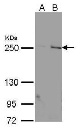
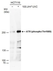
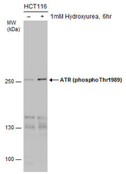
- ATR (phospho Thr1989) antibody detects ATR (phospho Thr1989) protein by Western blot analysis.A. 30 µg HCT116 whole cell lysate/extract (untreat)B. 30 µg HCT116 whole cell lysate/extract (100 J/m2 UVC treatment and recover for 6 hr) 5 % SDS-PAGEATR (phospho Thr1989) antibody (GTX128145) dilution: 1:1000
- Validation comment
- WB

- Submitted by
- GeneTex (provider)
- Main image
- Experimental details
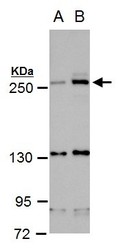
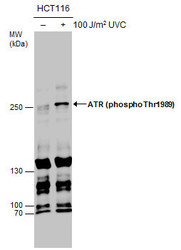
- ATR (phospho Thr1989) antibody detects ATR (phospho Thr1989) protein by western blot analysis.A. 30 ?g HCT116 whole cell extract (untreated)B. 30 ?g HCT116 whole cell extract (100 J/m2 UVC treatment and recover for 6 hr)5 % SDS-PAGEATR (phospho Thr1989) antibody (GTX128145) dilution: 1:1000
- Validation comment
- WB

- Submitted by
- GeneTex (provider)
- Main image
- Experimental details
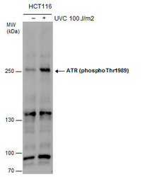
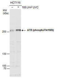
- ATR (phospho Thr1989) antibody detects ATR (phospho Thr1989) protein by western blot analysis. Un-treated (-) and treated (+, 100 J/m2 UVC treatment and recover for 6 hr) HCT116 whole cell extracts (30 ?g) were separated by 5% SDS-PAGE, and the membrane was blotted with ATR (phospho Thr1989) antibody (GTX128145) at a dilution of 1:1000.
- Validation comment
- WB

- Submitted by
- GeneTex (provider)
- Main image
- Experimental details
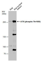
- Raji whole cell and nuclear extracts (30 ?g) were separated by 5% SDS-PAGE, and the membrane was blotted with ATR (phospho Thr1989) antibody (GTX128145) diluted at 1:500. The HRP-conjugated anti-rabbit IgG antibody (GTX213110-01) was used to detect the primary antibody.

- Submitted by
- GeneTex (provider)
- Main image
- Experimental details
- Untreated (¡V) and treated (+) HCT116 whole cell extracts (30 ?g) were separated by 5% SDS-PAGE, and the membrane was blotted with ATR (phospho Thr1989) antibody (GTX128145) diluted at 1:1000.

- Submitted by
- GeneTex (provider)
- Main image
- Experimental details
- Untreated (¡V) and treated (+) HCT116 whole cell extracts (30 £gg) were separated by 5% SDS-PAGE, and the membrane was blotted with ATR (phospho Thr1989) antibody (GTX128145) diluted at 1:1000.

- Submitted by
- GeneTex (provider)
- Main image
- Experimental details
- Untreated (¡V) and treated (+) HCT116 whole cell extracts (30 ?g) were separated by 5% SDS-PAGE, and the membrane was blotted with ATR (phospho Thr1989) antibody (GTX128145) diluted at 1:1000. The HRP-conjugated anti-rabbit IgG antibody (GTX213110-01) was used to detect the primary antibody.

- Submitted by
- GeneTex (provider)
- Main image
- Experimental details
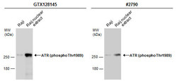
- Raji whole cell and nuclear extracts (30 ?g) were separated by 5% SDS-PAGE, and the membranes were blotted with ATR (phospho Thr1989) antibody (GTX128145) diluted at 1:500 and competitor's antibody (CST#2790) diluted at 1:1000. The HRP-conjugated anti-rabbit IgG antibody (GTX213110-01) was used to detect the primary antibody.

- Submitted by
- GeneTex (provider)
- Main image

- Experimental details
- Raji whole cell and nuclear extracts (30 ?g) were separated by 5% SDS-PAGE, and the membranes were blotted with ATR (phospho Thr1989) antibody (GTX128145) diluted at 1:500 and competitor's antibody (CST#2790) diluted at 1:1000. The HRP-conjugated anti-rabbit IgG antibody (GTX213110-01) was used to detect the primary antibody.

- Submitted by
- GeneTex (provider)
- Main image
- Experimental details
- Untreated (¡V) and treated (+) HCT-116 whole cell extracts (30 £gg) were separated by 5% SDS-PAGE, and the membrane was blotted with ATR (phospho Thr1989) antibody (GTX128145) diluted at 1:1000.

Supportive validation
- Submitted by
- GeneTex (provider)
- Main image

- Experimental details
- ATR (phospho Thr1989) antibody detects ATR (phospho Thr1989) protein at cytoplasm and nucleus by immunofluorescent analysis.Sample: HeLa cells were fixed in 4% paraformaldehyde at RT for 15 min.Green: ATR (phospho Thr1989) protein stained by ATR (phospho Thr1989) antibody (GTX128145) diluted at 1:500.Red: phalloidin, a cytoskeleton marker, diluted at 1:50.Scale bar = 10 £gm.

Supportive validation
- Submitted by
- GeneTex (provider)
- Main image

- Experimental details
- Immunoprecipitation of ATR (phospho Thr1989) protein from HCT-116 whole cell extracts treated with Hydroxyurea for 6 hr using 5 £gg of ATR (phospho Thr1989) antibody (GTX128145).Western blot analysis was performed using ATR (phospho Thr1989) antibody (GTX128145).EasyBlot anti-Rabbit IgG (GTX221666-01) was used as a secondary reagent.

Supportive validation
- Submitted by
- GeneTex (provider)
- Main image

- Experimental details
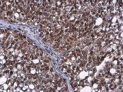
- ATR (phospho Thr1989) antibody detects ATR (phospho Thr1989) protein at nucleus on human ovarian carcinoma by immunohistochemical analysis. Sample: Paraffin-embedded human ovarian carcinoma. ATR (phospho Thr1989) antibody (GTX128145) diluted at 1:250.

- Submitted by
- GeneTex (provider)
- Main image

- Experimental details
- ATR (phospho Thr1989) antibody detects ATR (phospho Thr1989) protein at nucleus in human lung by immunohistochemical analysis. Sample: Paraffin-embedded human lung. ATR (phospho Thr1989) antibody (GTX128145) diluted at 1:250.

- Submitted by
- GeneTex (provider)
- Main image

- Experimental details
- ATR (phospho Thr1989) antibody detects ATR (phospho Thr1989) protein at nucleus in mouse liver by immunohistochemical analysis. Sample: Paraffin-embedded mouse liver. ATR (phospho Thr1989) antibody (GTX128145) diluted at 1:200.

- Submitted by
- GeneTex (provider)
- Main image
- Experimental details
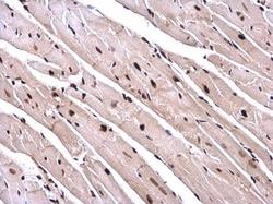
- ATR (phospho Thr1989) antibody detects ATR (phospho Thr1989) protein at nucleus in mouse heart by immunohistochemical analysis. Sample: Paraffin-embedded mouse heart. ATR (phospho Thr1989) antibody (GTX128145) diluted at 1:200.

- Submitted by
- GeneTex (provider)
- Main image

- Experimental details
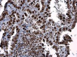
- ATR (phospho Thr1989) antibody detects ATR protein at nucleus in human lung cancer by immunohistochemical analysis. Sample: Paraffin-embedded human lung cancer. ATR (phospho Thr1989) antibody (GTX128145) diluted at 1:250.

- Submitted by
- GeneTex (provider)
- Main image

- Experimental details
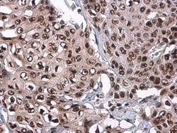
- ATR (phospho Thr1989) antibody detects ATR (phospho Thr1989) protein at nucleus in human breast carcinoma by immunohistochemical analysis. Sample: Paraffin-embedded human breast carcinoma. ATR (phospho Thr1989) antibody (GTX128145) diluted at 1:250.

- Submitted by
- GeneTex (provider)
- Main image
- Experimental details
- ATR (phospho Thr1989) antibody detects ATR (phospho Thr1989) protein at nucleus in human colon cancer by immunohistochemical analysis. Sample: Paraffin-embedded human colon cancer. ATR (phospho Thr1989) antibody (GTX128145) diluted at 1:250.

- Submitted by
- GeneTex (provider)
- Main image
- Experimental details
- ATR (phospho Thr1989) antibody detects ATR (phospho Thr1989) protein at nucleus by immunohistochemical analysis.Sample: Paraffin-embedded human esophageal carcinoma.ATR (phospho Thr1989) stained by ATR (phospho Thr1989) antibody (GTX128145) diluted at 1:500.

- Submitted by
- GeneTex (provider)
- Main image
- Experimental details
- ATR (phospho Thr1989) antibody detects ATR (phospho Thr1989) protein at nucleus by immunohistochemical analysis.Sample: Paraffin-embedded human ovarian cancer.ATR (phospho Thr1989) stained by ATR (phospho Thr1989) antibody (GTX128145) diluted at 1:500.
